May. 07, 2020
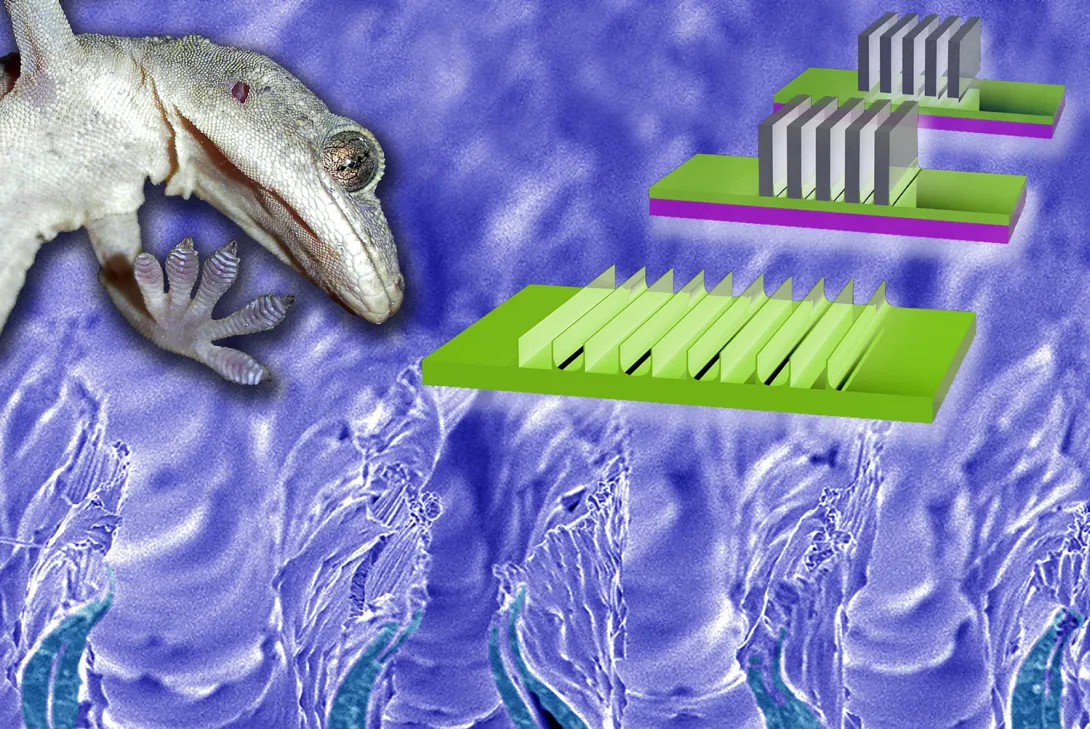
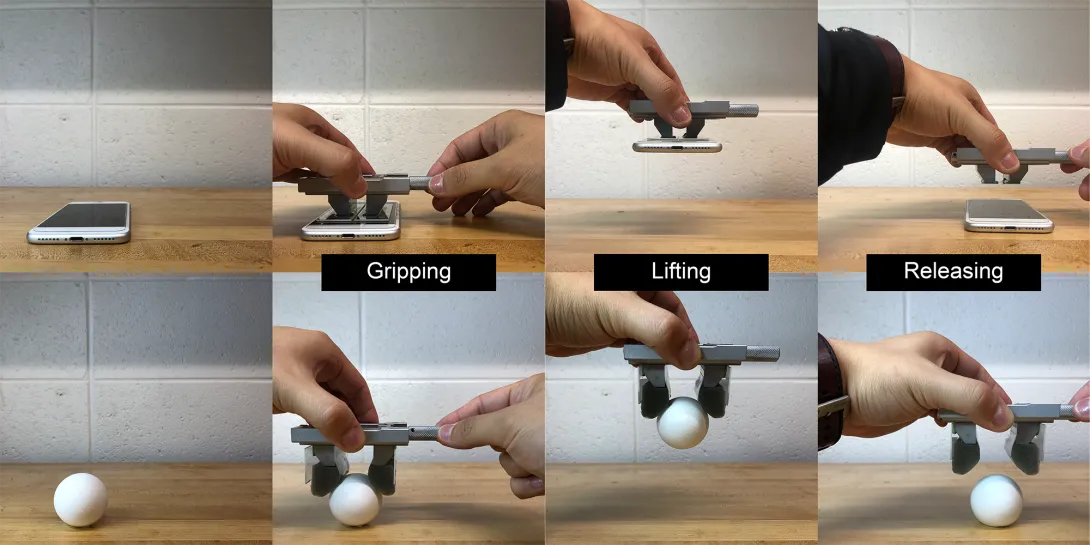
Why did the gecko climb the skyscraper? Because it could; its toes stick to about anything. Engineers can already emulate the secrets of gecko stickiness to make strips of rubbery materials that can pick up and release objects, but simple mass production for everyday use has been out of reach until now.
Researchers at the Georgia Institute of Technology have developed, in a new study, a method of making gecko-inspired adhesive materials that is much more cost-effective than current methods. It could enable mass production and the spread of the versatile gripping strips to manufacturing and homes.
Polymers with “gecko adhesion” surfaces could be used to make extremely versatile grippers to pick up very different objects even on the same assembly line. They could make picture hanging easy by adhering to both the picture and the wall at the same time. Vacuum cleaner robots with gecko adhesion could someday scoot up tall buildings to clean facades.
“With the exception of things like Teflon, it will adhere to anything. This is a clear advantage in manufacturing because we don’t have to prepare the gripper for specific surfaces we want to lift. Gecko-inspired adhesives can lift flat objects like boxes then turn around and lift curved objects like eggs and vegetables,” said Michael Varenberg, the study’s principal investigator and an assistant professor in Georgia Tech’s George W. Woodruff School of Mechanical Engineering.
Current grippers on assembly lines, such as clamps, magnets, and suction cups, can each lift limited ranges of objects. Grippers based on gecko-inspired surfaces, which are dry and contain no glue or goo, could replace many grippers or just fill in capability gaps left by other gripping mechanisms.
Drawing out razors
The adhesion comes from protrusions a few hundred microns in size that often look like sections of short, floppy walls running parallel to each other across the material’s surface. How they work by mimicking geckos’ feet is explained below.
Up to now, molding has produced these mesoscale walls by pouring ingredients onto a template, letting the mixture react and set to a flexible polymer then removing it from the mold. But the method is inconvenient.
“Molding techniques are expensive and time-consuming processes. And there are issues with getting the gecko-like material to release from the template, which can disturb the quality of the attachment surface,” Varenberg said.
The researchers’ new method formed those walls by pouring ingredients onto a smooth surface instead of a mold, letting the polymer partially set then dipping rows of laboratory razor blades into it. The material set a little more around the blades, which were then drawn out, leaving behind micron-scale indentations surrounded by the desired walls.
Varenberg and first author Jae-Kang Kim published details of their new method in the journal ACS Applied Materials & Interfaces on April 6, 2020.
Forget about perfection
Though the new method is easier than molding, developing it took a year of dipping, drawing, and readjusting while surveying finicky details under an electron microscope.
“There are many parameters to control: Viscosity and temperature of the liquid; timing, speed, and distance of withdrawing the blades. We needed enough plasticity of the setting polymer to the blades to stretch the walls up, and not so much rigidity that would lead the walls to rip up,” Varenberg said.
Gecko-inspired surfaces have a fine topography on a micron-scale and sometimes even on a nanoscale, and surfaces made via molding are usually the most precise. But such perfection is unnecessary; the materials made with the new method did the job well and were also markedly robust.
“Many researchers demonstrating gecko adhesion have to do it in a cleanroom in clean gear. Our system just plain works in normal settings. It is robust and simple, and I think it has good potential for use in industry and homes,” said Varenberg, who studies surfaces in nature to mimic their advantageous qualities in human-made materials.
[Ready for graduate school with social distancing? Here's how to apply to Georgia Tech.]
Gecko foot fluff
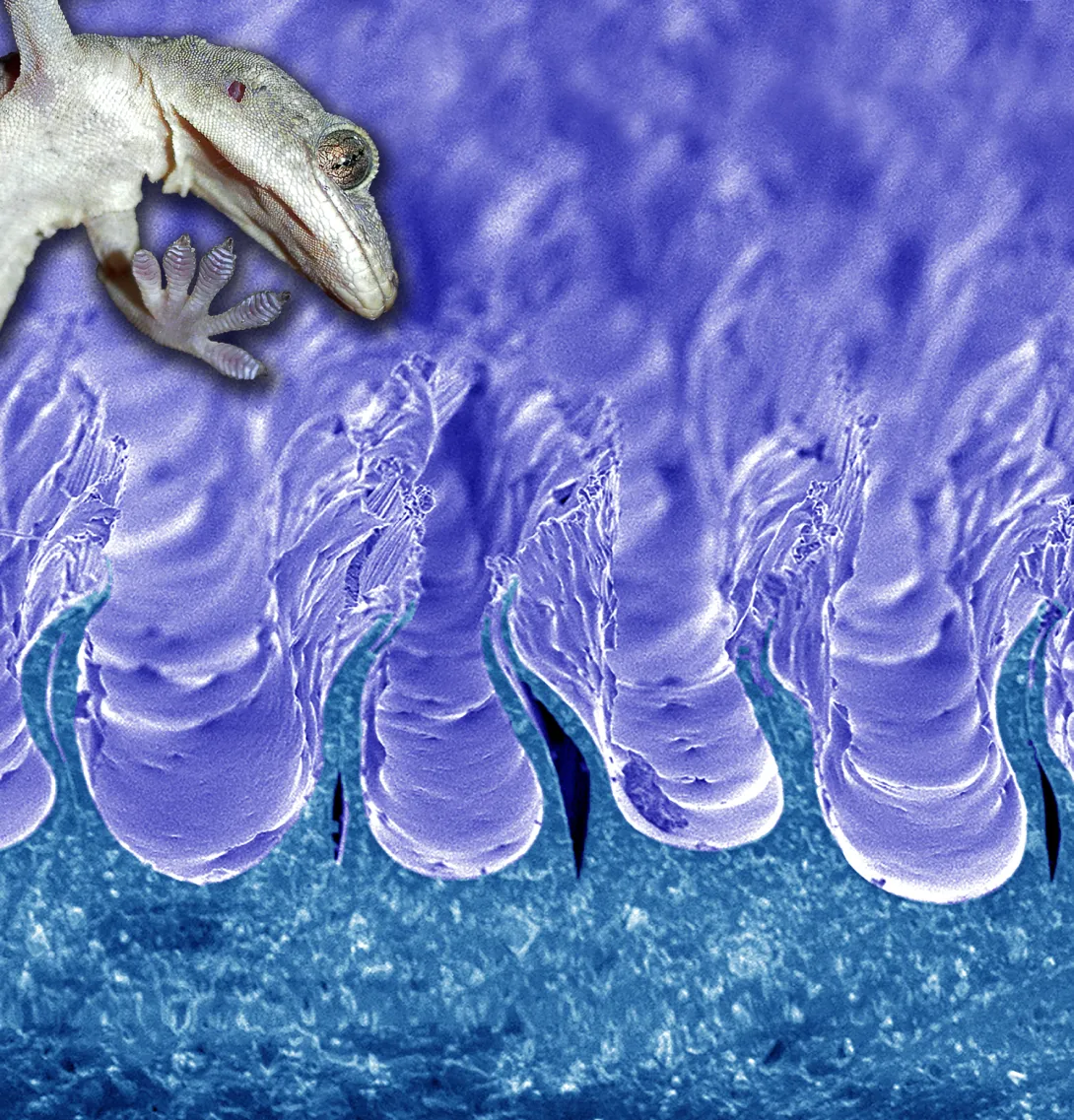
Behold the gecko’s foot. It has ridges on its toes, and this has led some in the past to think their feet stick by suction or some kind of clutching by the skin.
But electron microscopes reveal a deeper structure – spatula-shaped bristly fibrils protrude a few dozen microns long off those ridges. The fibrils make such thorough contact with surfaces down to the nanoscale that weak attractions between atoms on both sides appear to add up enormously to create overall strong adhesion.
In place of fluff, engineers have developed rows of shapes covering materials that produce the effect. A common shape makes a material’s surface look like a field of mushrooms that are a few hundred microns in size; another is rows of short walls like those in this study.
“The mushroom patterns touch a surface, and they are attached straightaway, but detaching requires applying forces that can be disadvantageous. The wall-shaped projections require minor shear force like a tug or a gentle grab to generate adherence, but that is easy, and letting go of the object is uncomplicated, too,” Varenberg said.
Varenberg’s research team used the drawing method to make walls with U-shaped spaces in between them and walls with V-shaped spaces in between. They worked with polyvinylsiloxane (PVS) and polyurethane (PU). The V-shape made in PVS worked best, but polyurethane is the better material for industry, so Vanenberg’s group will now work toward achieving the V-shape gecko gripping pattern in PU for the best possible combination.
Also read: Lung-heart super sensor on a chip tinier than a ladybug
Here's how to subscribe to our free science and technology email newsletter
Writer & Media Representative: Ben Brumfield (404-272-2780), email: ben.brumfield@comm.gatech.edu
Georgia Institute of Technology
Apr. 22, 2020

Gaps in the supply of coronavirus tests are propelling initiatives to fill them across the country. At the Georgia Institute of Technology, bioscience researchers are burning the midnight oil to produce key components for tests in the state of Georgia.
The goal is to supply a broad initiative by the governor’s office involving multiple universities and partners to rapidly produce and administer more tests. At least 35 volunteers at Georgia Tech, while adhering to social distancing, are reorienting labs normally used for scientific discovery to do larger-scale production of biochemical components.
“We are inventing new ways of doing things like an electronic buddy system so people can be alone – but not alone – while they work in the lab. The technical part is actually the easiest. The logistics of testing, data security, and regulatory considerations – those things are more challenging,” said Loren Williams, a professor in Georgia Tech’s School of Chemistry and Biochemistry.
Williams and the researchers are supporting Georgia Governor Brian Kemp’s COVID-19 State Lab Surge Capacity Task Force, which is a project managed through the Georgia Tech Research Institute (GTRI). GTRI is also leading the coordination and integration of data management across the lab surge effort.
“We are providing technical and project management of the effort which is focused on increasing the state’s ability to expand testing beyond current limitations,” said Mike Shannon, GTRI’s lead in the project and a principal research engineer at GTRI.
Exoplanets and coronavirus
The science behind coronavirus testing is complementary to the researchers’ usual work. That includes understanding proteins associated with glaucoma, figuring out how RNA and DNA evolved in the first place, or whether ribosomes – lumps of RNA and protein key to translating genetic code into life – may exist on exoplanets.
Williams’ research team studies the last topic, and some of their work is related to the core of coronavirus testing, a chemical reaction that amplifies the virus’ genetic fingerprint. It is called a reverse transcription polymerase chain reaction (RT-PCR), and it transcribes trace amounts of coronavirus’ RNA code into ample amounts of corresponding DNA in the lab for easy analysis.
“His lab members are very familiar with RT-PCR, and when the lack of tests became apparent, they swung into action. The group grew from there, based on the technical needs for the project,” said Raquel Lieberman, another leading scientist in the effort and also a professor in Georgia Tech’s School of Chemistry and Biochemistry.
“Every day, very talented, hardworking people with perfect skill sets come out of the woodwork and ask to help,” Williams said.
The group has teams that engineer the production of enzymes or other chemicals needed for RT-PCR to work: Two central enzymes are reverse transcriptase, which converts RNA to DNA and Taq polymerase, which rapidly replicates DNA. Another important component is ribonuclease inhibitor, which slows coronavirus RNA decay.
Global COVID allies
Other researchers develop processes for mass production or implementation of COVID-19 safety procedures; the list goes on. Some colleagues telework; others work in labs but spaced far from each other while they wear masks.
“The group is planning to produce enough enzyme components for hundreds of tests per day,” said Vinayah Agarwal, an assistant professor in Georgia Tech’s School of Chemistry and Biochemistry and School of Biological Sciences. “Using these components, we will also build cheaper and more robust testing kits going forward.”
Instructions already exist for some of the ingredients for the test, but they are not readily available because the rights to them are exclusive.
“Intellectual property and other proprietary issues hinder our effort,” Lieberman said. “But we have received help from scientists all over the world to piece together protocols on how to make what we need.”
The state wants to increase current testing capacities by 3,000 more tests per day. The task force also includes teams from Augusta University Health System, Georgia State University, Emory University, University of Georgia, and the Georgia Public Health Laboratory. The task force lead is Captain Kevin Caspary who is with the Georgia National Guard.
Raw footage and images as press handouts for journalists. (No commercial or personal use):
https://www.dropbox.com/sh/f2wc2i74lz1lffl/AADLJ8dQnZMr4uEDxAiIMusoa?dl=0
Also read this: Interactive COVID-19 tool shows the importance of staying at home
External News Coverage:
NPR - Sun Rays, Disinfectants And False Hopes: Misinformation Litters The Road To Reopening
News-Medical.Net - Georgia Tech researchers create key components for COVID-19 tests
Georgia Tech News Center- A New Normal: Researchers Across Georgia Tech Rally to Fight COVID-19
Here's how to subscribe to our free science and technology email newsletter
Writer & Media Representative: Ben Brumfield (404-272-2780), email: ben.brumfield@comm.gatech.edu
Georgia Institute of Technology
Apr. 15, 2020

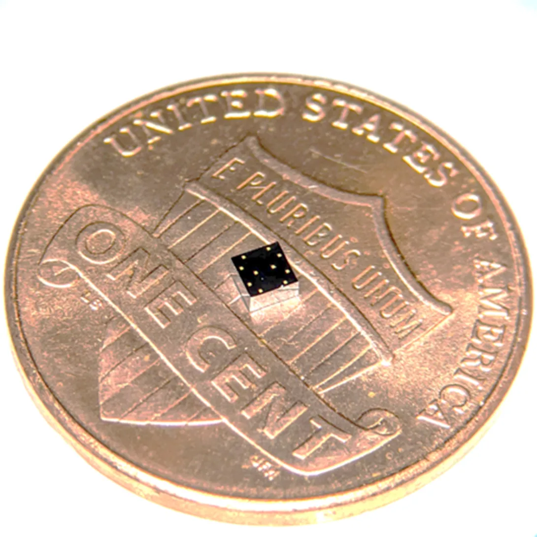
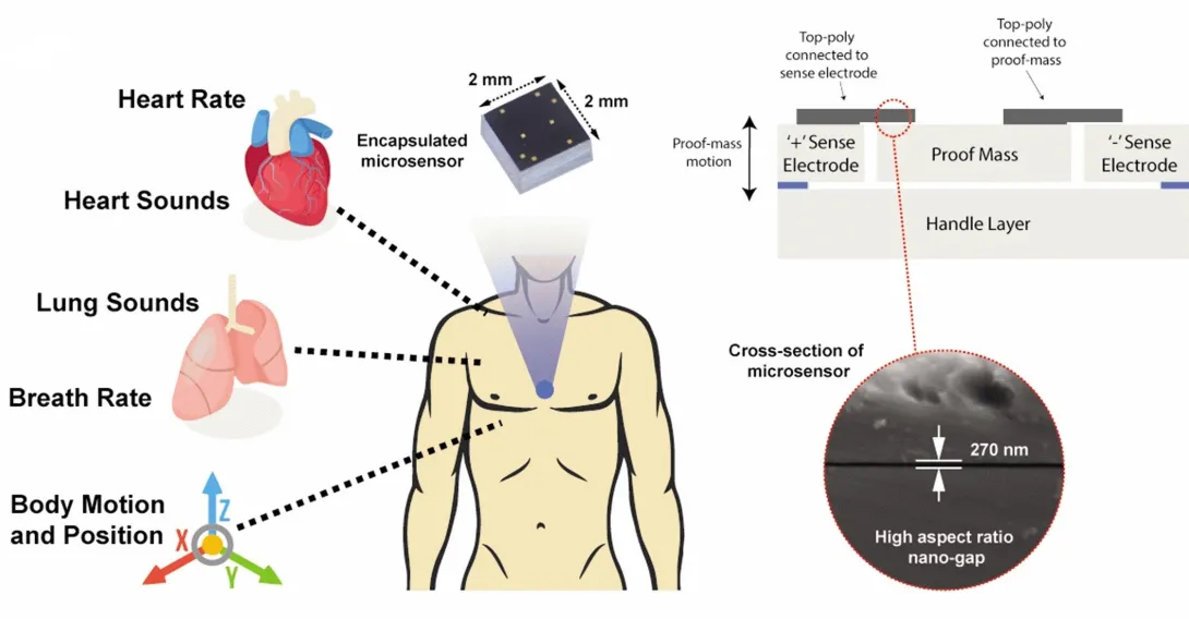
During a stroll, a woman’s breathing becomes a slight bit shallower, and a monitor in her clothing alerts her to get a telemedicine check-up. A new study details how a sensor chip smaller than a ladybug records multiple lung and heart signals along with body movements and could enable such a future socially distanced health monitor.
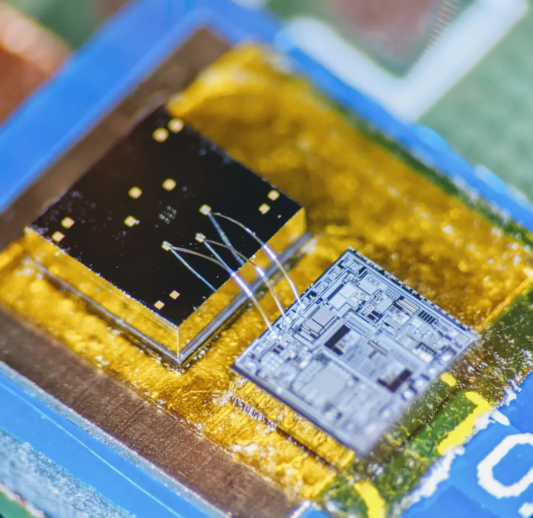
The core mechanism of the chip developed by researchers at the Georgia Institute of Technology involves two finely manufactured layers of silicon, which overlay each other separated by the space of 270 nanometers – about 0.000001 inches. They carry a minute voltage.
Vibrations from bodily motions and sounds put part of the chip in very slight motion, making the voltage flux, thus creating readable electronic outputs. In human testing, the chip has recorded a variety of signals from the mechanical workings of the lungs and the heart with clarity, signals that often escape meaningful detection by current medical technology.
“Right now, medicine looks to EKGs (electrocardiograms) for information on the heart, but EKGs only measure electrical impulses. The heart is a mechanical system with muscles pumping and valves opening and shutting, and it sends out a signature of sounds and motions, which an EKG does not detect. EKGs also say nothing about lung function,” said Farrokh Ayazi, Ken Byers Professor in Georgia Tech’s School of Electrical and Computer Engineering.
Stethoscope-accelerometer combo
The chip, which acts as an advanced electronic stethoscope and accelerometer in one, is aptly called an accelerometer contact microphone. It detects vibrations that enter the chip from inside the body while keeping out distracting noise from outside the body's core like airborne sounds
“If it rubs on my skin or shirt, it doesn’t hear the friction, but the device is very sensitive to sounds coming at it from inside the body, so it picks up useful vibrations even through clothing,” Ayazi said.
The detection bandwidth is enormous - from broad, sweeping motions to inaudibly high-pitched tones. Thus, the sensor chip records all at once fine details of the heartbeat, waves the heart sends through the body, and respiration rates and lung sounds. It even tracks the wearer’s physical activities such as walking.
The signals are recorded in sync, potentially offering the big picture of a patient’s heart and lung health. For the study, the researchers successfully recorded a “gallop,” a faint third sound after the “lub-dub” of the heartbeat. Gallops are normally elusive clues of heart failure.
The researchers published their results in the journal npj Digital Medicine on February 12, 2020. The research was funded by the Georgia Research Alliance, the Defense Advanced Research Projects Agency (DARPA), the National Science Foundation, and the National Institutes of Health. Study coauthor Divya Gupta, M.D., a cardiologist at Emory University, collaborated in testing the chip on human participants.
Hermetically sealed vacuum
Medical research has tried to make better use of the body’s mechanical signals for decades but recording some – like waves traversing multiple tissues – has proven inconsistent, while others – like gallops – have relied upon clinician skills influenced by human error. The new chip produces high-resolution, quantified data that future research could match to pathologies in order to identify them.
“We are working already to collect significantly more data matched with pathologies. We envision algorithms in the future that may enable a broad array of clinical readings,” Ayazi said.
Though the chip’s main engineering principle is simple, making it work and then manufacturable took Ayazi’s lab ten years, mainly because of the Lilliputian scale of the gap between the silicon layers, i.e. electrodes. If the 2-millimeter by 2-millimeter sensor chip were expanded to the size of a football field, that air gap would be about an inch wide.
“That very thin gap separating the two electrodes cannot have any contact, not even by forces in the air in between the layers, so the whole sensor is hermetically sealed inside a vacuum cavity,” Ayazi said. “This makes for that ultralow signal noise and breadth of bandwidth that are unique.”
Detects through clothing
The researchers used a manufacturing process developed in Ayazi’s lab called the HARPSS+ platform (High Aspect Ratio Poly and Single Crystalline Silicon) for mass production, running off hand-sized sheets that were then cut into the tiny sensor chips. HARPSS+ is the first reported mass manufacturing process that achieves such consistently thin gaps, and it has enabled high-throughput manufacturing of many such advanced MEMS, or microelectromechanical systems.
The experimental device is currently battery-powered and uses a second chip called a signal-conditioning circuit to translate the sensor chip’s signals into patterned read-outs.
Three sensors or more could be inserted into a chest band that would triangulate health signals to locate their sources. Someday a device may pinpoint an emerging heart valve flaw by turbulence it produces in the bloodstream or identify a cancerous lesion by faint crackling sounds in a lung.
Here's how to subscribe to our free science and technology newsletter
Also read: Digital tool helps with tough COVID19 decision
These researchers co-authored the study: Pranav Gupta (first author), Mohammad Moghimi, Yaesuk Jeong and Omer Inan from Georgia Tech. The research was funded by the Georgia Research Alliance, the Defense Advanced Research Projects Agency (DARPA) Technology Office’s Advanced Inertial Micro Sensors program (contract # N66001-16-1-4064), and by the National Science Foundation/National Institutes of Health Smart and Connected Health Program (grant # R01 EB023808). The team’s work with human subjects was approved by Emory University and Georgia Institute of Technology Institutional Review Boards (IRB# H18248). Any findings, conclusions or recommendations are those of the authors and not necessarily of the sponsors.
Writer & Media Representative: Ben Brumfield (404-272-2780), email: ben.brumfield@comm.gatech.edu
Georgia Institute of Technology
Apr. 13, 2020


What-if questions can torment a doctor making coronavirus retest decisions: What if a patient’s initial negative test was a false negative, and he or she needs a second test? What if they don’t need it, and a retest would use up a scarce test kit and treatments that other patients need?
Such challenges led Piedmont Healthcare in Atlanta to establish a paper-based decision tree for ordering COVID-19 retests, and researchers at the Georgia Institute of Technology turned it into an automated digital tool. Piedmont further developed the tool and has now built it into the hospital’s electronic medical record, where it influences the ordering of retests.
A user can answer their “ifs” by clicking through questions, and the “if-this-then-do-that” algorithm makes recommendations for best courses of action, ranging from immediately treating a patient for COVID-19 to retesting to consulting a specialist. The final decision remains with the physician.
The questions are deceptively simple, but the recommendations are not always obvious. That reflects the algorithm’s usefulness to fill gaps in thinking about the new sickness, which can confront clinicians with surprises.
“If a patient has not had close contact with positive patients and the first test came back negative, a physician may think the patient does not need to be retested. But actually, the patient may need a second test because they are in intensive care and also have suspicious chest X-rays,” said Georgia Tech graduate research assistant April Yu, who converted the decision tree into a digital tool.
“One of our big worries in using a brand-new test like the coronavirus test is that it will miss real cases, and this tool helps prevent that,” said Dr. Bronwen Garner, who helped develop the original decision tree and is an infectious disease specialist at Piedmont Healthcare. “It also helps reassure physicians when they get a negative result that it is probably a true negative.”
Suspenseful decision-making
A physician’s reaction to an initial negative test can mean life or death because the physician not only decides on follow-up testing but also on treatment pathways and quarantine.
“If you make a misstep in the thought process, it can lead to cascading impacts not only for the patient but also for healthcare professionals and family members, who may be exposed to the patient,” said Pinar Keskinocak, William W. George Chair and Professor in Georgia Tech’s Stewart School of Industrial and Systems Engineering. “This tool is meant to help doctors easily stay on the decision tree path.”
Michael O’Toole, executive director of Piedmont Healthcare’s quality improvement department, originally pictured doctors getting an automated version of the decision tree to use on their phones. O’Toole called Keskinocak, and she tapped Yu, a member of her research group.
“Literally within four hours they had it ready for us. It was incredible,” said O’Toole, a Georgia Tech alumnus who studied industrial and systems engineering.
“It was a very pleasant surprise,” said Dr. Garner, who is also a Georgia Tech graduate. “Automated tools are better than a paper format because they’re in the same format as orders in our electronic system. We get notifications in real time instead of having to remember to check a piece of paper.”
The tool is in place in the system where doctors order retests and is specific to Piedmont’s workflow. It may not be directly transferable to other health care systems.
Piedmont Healthcare simplified the logic even more, and the hospital built its own custom alerts to guide physicians on retesting. For cases that are more ambiguous, Piedmont Healthcare’s final version of the tool also gives physicians inside the hospital guidance to consult with their in-house infectious disease specialists.
If-this-then-retest
In her original version, Yu had turned the decision tree criteria into a short panel of questions with yes and no answers. It took her six iterations to arrive at her final version.
Yu’s version asked whether the patient:
- has a relevant ailment
- previously tested positive for coronavirus
- is now in an intensive care unit
- has worsening lung conditions
- shows telltale lung damage in imaging
- has been diagnosed with a different ailment
- the patient has had contact with someone else who tested positive for coronavirus.
On the back end, the algorithm guided the user through risks of coronavirus presence based on the answers.
“The steps were easy to follow, and the answers were color-coded for urgency with white, yellow, and red,” said Keskinocak, who also directs Georgia Tech’s Center for Health and Humanitarian Systems.
One bright yellow answer read: “This patient needs re-testing 24 hours after the initial test!” And there were further recommendations on how to handle the case.
Here's how to subscribe to our free science and technology newsletter
Also read: Advice on DIY masks
Writer & Media Representative: Ben Brumfield (404-272-2780), email: ben.brumfield@comm.gatech.edu
Georgia Institute of Technology
Feb. 11, 2020

Four Georgia Institute of Technology faculty members have been elected as new members of the National Academy of Engineering (NAE). Marilyn Brown, Thomas Kurfess, Susan Margulies, and Alexander Shapiro join 83 other new NAE members for 2020 when they are formally inducted during a ceremony at the academy’s annual meeting on Oct. 4 in Washington, D.C.
Election of new NAE members, the culmination of a yearlong process, recognizes individuals who have made outstanding contributions to "engineering research, practice, or education, including, where appropriate, significant contributions to the engineering literature" and to "the pioneering of new and developing fields of technology, making major advancements in traditional fields of engineering, or developing/implementing innovative approaches to engineering education."
“It’s the honor of a lifetime to be recognized by the National Academy of Engineering for the impact we’ve have on understanding lung injuries in the critical care unit and traumatic brain injuries in children,” said Margulies, chair of the Wallace H. Coulter Department of Biomedical Engineering at Georgia Tech and Emory University and, with Brown, one of just three women on the Georgia Tech faculty accorded NAE membership – one of the highest professional distinctions an engineer can receive.
“Our work is deeply collaborative, and I am grateful to the engineers, scientists, physicians, and patients who are partners in our journey,” Margulies added.
Margulies, a researcher in the Petit Institute for Bioengineering and Bioscience at Tech and a Georgia Research Alliance Eminent Scholar in Injury Biomechanics at Emory, was elected, “for elaborating the traumatic injury thresholds of brain and lung in terms of structure-function mechanisms,” according to the NAE announcement.
Using an integrated biomechanics approach, Margulies’ research program spans the micro-to-macro scales in two distinct areas, traumatic brain injury and ventilator-induced lung injury. Her work has generated new knowledge about the structural and functional responses of the brain and lungs to their mechanical environment. Margulies came to Georgia Tech in 2017 from the University of Pennsylvania, where she’d been a professor of bioengineering, and had earned her Master of Science in Engineering and Ph.D. in Bioengineering.
Brown, a Regents and Brook Byers Professor of Sustainable Systems in the School of Public Policy, was co-recipient of the Nobel Peace Prize in 2007 (for co-authorship of the Intergovernmental Panel on Climate Change Working Group III Assessment Report on Mitigation of Climate Change, Chapter 6).
She joined Georgia Tech in 2006 after a career at the U.S. Department of Energy's Oak Ridge National Laboratory, where she led several national climate change mitigation studies and became a leader in the analysis and interpretation of energy futures in the United States. Her research at Tech focuses on the design and impact of policies aimed at accelerating the development and deployment of sustainable energy technologies, emphasizing the electric utility industry. She was elected to NAE “for bridging engineering, social and behavioral sciences, and policy studies to achieve cleaner electric energy.”
Brown, who earned her Ph.D. at the Ohio State University, co-founded and chaired the Southeast Energy Efficiency Alliance, served two terms as a presidential appointee on the board of the Tennessee Valley Authority – the nation’s largest public power provider – and also served two terms on the U.S. Department of Energy’s Electricity Advisory Committee, where she led the Smart Grid Subcommittee.
“The most rewarding feature of my career has been working toward solutions with colleagues across disciplines,” Brown said.
Shapiro is the Russell Chandler III Chair and professor in the H. Milton Stewart School of Industrial and Systems Engineering, where his research is focused on stochastic programming, risk analysis, simulation-based optimization, and multivariate statistical analysis.
In 2013, he was awarded the INFORMS Khachiyan Prize for lifetime achievements in optimization. He received the 2018 Dantzig Prize from the Mathematical Optimization Society and the Society for Industrial and Applied Mathematics.
Since earning his Ph.D. in applied mathematics-statistics from Israel’s Ben-Gurion University of the Negev in 1981, Shapiro has made substantial contributions to the fields of optimization and large-scale, stochastic programming, and he was elected to NAE “for contributions to the theory, computation, and application of stochastic programming.”
Kurfess is professor and HUSCO/Ramirez Distinguished Chair in Fluid Power and Motion Control in the George W. Woodruff School of Mechanical Engineering, where he has helped guide the evolution of technology as a pioneer in the digital transformation of manufacturing.
Improving manufacturing technology is a pursuit that has roots in his childhood. “I grew up in my father’s machine shop,” said Kurfess, who has a special fondness for mom-and-pop operations. He was elected by the NAE “for development and implementation of innovative digital manufacturing technologies and system architectures.”
“I’m proud that the work we do has a positive impact on small and medium-sized enterprises, which are about 99% of the manufacturing operations, as well as large operations,” said Kurfess, who earned all of his degrees at MIT. “Our work targets people who are implementing the digital thread in manufacturing, and what the digital thread will do is make sure those smaller enterprises, those mom and pops, can have access to the latest and greatest technologies.”
Research News
Georgia Institute of Technology
177 North Avenue
Atlanta, Georgia 30332-0181 USA
Media Relations Contact: John Toon (404-894-6986) (jtoon@gatech.edu).
Writer: Jerry Grillo
News Contact
John Toon
Research News
(404) 894-6986
May. 10, 2012
New research findings show that embryonic stem cells unable to fully compact the DNA inside them cannot complete their primary task: differentiation into specific cell types that give rise to the various types of tissues and structures in the body.
Researchers from the Georgia Institute of Technology and Emory University found that chromatin compaction is required for proper embryonic stem cell differentiation to occur. Chromatin, which is composed of histone proteins and DNA, packages DNA into a smaller volume so that it fits inside a cell.
A study published on May 10, 2012 in the journal PLoS Genetics found that embryonic stem cells lacking several histone H1 subtypes and exhibiting reduced chromatin compaction suffered from impaired differentiation under multiple scenarios and demonstrated inefficiency in silencing genes that must be suppressed to induce differentiation.
“While researchers have observed that embryonic stem cells exhibit a relaxed, open chromatin structure and differentiated cells exhibit a compact chromatin structure, our study is the first to show that this compaction is not a mere consequence of the differentiation process but is instead a necessity for differentiation to proceed normally,” said Yuhong Fan, an assistant professor in the Georgia Tech School of Biology.
Fan and Todd McDevitt, an associate professor in the Wallace H. Coulter Department of Biomedical Engineering at Georgia Tech and Emory University, led the study with assistance from Georgia Tech graduate students Yunzhe Zhang and Kaixiang Cao, research technician Marissa Cooke, and postdoctoral fellow Shiraj Panjwani.
The work was supported by the National Institutes of Health’s National Institute of General Medical Sciences (NIGMS), the National Science Foundation, a Georgia Cancer Coalition Distinguished Scholar Award, and a Johnson & Johnson/Georgia Tech Healthcare Innovation Award.
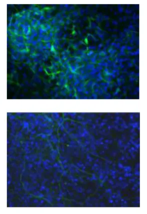
To investigate the impact of linker histones and chromatin folding on stem cell differentiation, the researchers used embryonic stem cells that lacked three subtypes of linker histone H1 -- H1c, H1d and H1e -- which is the structural protein that facilitates the folding of chromatin into a higher-order structure. They found that the expression levels of these H1 subtypes increased during embryonic stem cell differentiation, and embryonic stem cells lacking these H1s resisted spontaneous differentiation for a prolonged time, showed impairment during embryoid body differentiation and were unsuccessful in forming a high-quality network of neural cells.
“This study has uncovered a new, regulatory function for histone H1, a protein known mostly for its role as a structural component of chromosomes,” said Anthony Carter, who oversees epigenetics grants at NIGMS. “By showing that H1 plays a part in controlling genes that direct embryonic stem cell differentiation, the study expands our understanding of H1’s function and offers valuable new insights into the cellular processes that induce stem cells to change into specific cell types.”
During spontaneous differentiation, the majority of the H1 triple-knockout embryonic stem cells studied by the researchers retained a tightly packed colony structure typical of undifferentiated cells and expressed high levels of Oct4 for a prolonged time. Oct4 is a pluripotency gene that maintains an embryonic stem cell’s ability to self-renew and must be suppressed to induce differentiation.
“H1 depletion impaired the suppression of the Oct4 and Nanog pluripotency genes, suggesting a novel mechanistic link by which H1 and chromatin compaction may mediate pluripotent stem cell differentiation by contributing to the epigenetic silencing of pluripotency genes,” explained Fan. “While a significant reduction in H1 levels does not interfere with embryonic stem cell self-renewal, it appears to impair differentiation.”
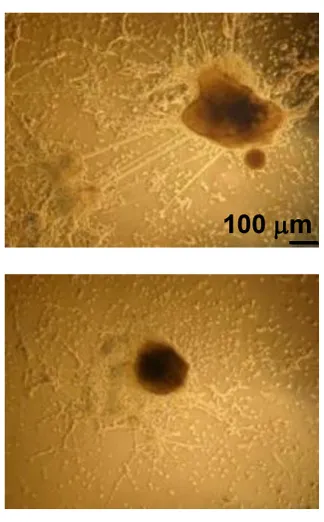
The researchers also used a rotary suspension culture method developed by McDevitt to produce with high efficiency homogonous 3D clumps of embryonic stem cells called embryoid bodies. Embryoid bodies typically contain cell types from all three germ layers -- the ectoderm, mesoderm and endoderm -- that give rise to the various types of tissues and structures in the body. However, the majority of the H1 triple-knockout embryoid bodies formed in rotary suspension culture lacked differentiated structures and displayed gene expression signatures characteristic of undifferentiated stem cells.
“H1 triple-knockout embryoid bodies displayed a reduced level of activation of many developmental genes and markers in rotary culture, suggesting that differentiation to all three germ layers was affected.” noted McDevitt.
The embryoid bodies also lacked the epigentic changes at the pluripotency genes necessary for differentiation, according to Fan.
“When we added one of the deleted H1 subtypes to the embryoid bodies, Oct4 was suppressed normally and embryoid body differentiation continued,” explained Fan. “The epigenetic regulation of Oct4 expression by H1 was also evident in mouse embryos.”
In another experiment, the researchers provided an environment that would encourage embryonic stem cells to differentiate into neural cells. However, the H1 triple-knockout cells were defective in forming neuronal and glial cells and a neural network, which is essential for nervous system development. Only 10 percent of the H1 triple-knockout embryoid bodies formed neurites and they produced on average eight neurites each. In contrast, half of the normal embryoid bodies produced, on average, 18 neurites.
In future work, the researchers plan to investigate whether controlling H1 histone levels can be used to influence the reprogramming of adult cells to obtain induced pluripotent stem cells, which are capable of differentiating into tissues in a way similar to embryonic stem cells.
Research reported in this publication was supported by the National Institute of General Medical Sciences of the National Institutes of Health (NIH) under award number GM085261 and the National Science Foundation under award number CBET-0939511. The content is solely the responsibility of the principal investigators and does not necessarily represent the official views of the NIH or NSF.
Research News & Publications Office
Georgia Institute of Technology
75 Fifth Street, N.W., Suite 314
Atlanta, Georgia 30308 USA
Media Relations Contacts: Abby Robinson (abby@innovate.gatech.edu; 404-385-3364) or John Toon (jtoon@gatech.edu; 404-894-6986)
Writer: Abby Robinson
News Contact
Abby Robinson
Research News and Publications
abby@innovate.gatech.edu
404-385-3364
May. 06, 2012
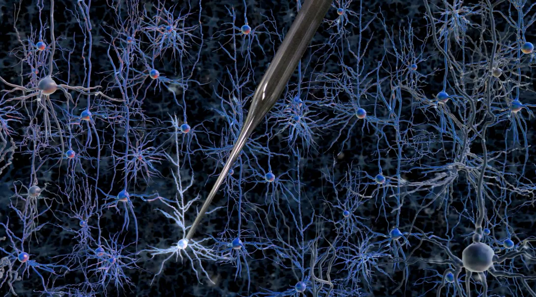
Gaining access to the inner workings of a neuron in the living brain offers a wealth of useful information: its patterns of electrical activity, its shape, even a profile of which genes are turned on at a given moment. However, achieving this entry is such a painstaking task that it is considered an art form; it is so difficult to learn that only a small number of labs in the world practice it.
But that could soon change: Researchers at MIT and the Georgia Institute of Technology have developed a way to automate the process of finding and recording information from neurons in the living brain. The researchers have shown that a robotic arm guided by a cell-detecting computer algorithm can identify and record from neurons in the living mouse brain with better accuracy and speed than a human experimenter.
The new automated process eliminates the need for months of training and provides long-sought information about living cells’ activities. Using this technique, scientists could classify the thousands of different types of cells in the brain, map how they connect to each other, and figure out how diseased cells differ from normal cells.
The project is a collaboration between the labs of Ed Boyden, associate professor of biological engineering and brain and cognitive sciences at MIT, and Craig Forest, an assistant professor in the George W. Woodruff School of Mechanical Engineering at Georgia Tech.
“Our team has been interdisciplinary from the beginning, and this has enabled us to bring the principles of precision machine design to bear upon the study of the living brain,” Forest says. His graduate student, Suhasa Kodandaramaiah, spent the past two years as a visiting student at MIT, and is the lead author of the study, which appears in the May 6 issue of Nature Methods.
The method could be particularly useful in studying brain disorders such as schizophrenia, Parkinson’s disease, autism and epilepsy, Boyden says. “In all these cases, a molecular description of a cell that is integrated with [its] electrical and circuit properties … has remained elusive,” says Boyden, who is a member of MIT’s Media Lab and McGovern Institute for Brain Research. “If we could really describe how diseases change molecules in specific cells within the living brain, it might enable better drug targets to be found.”
Automation
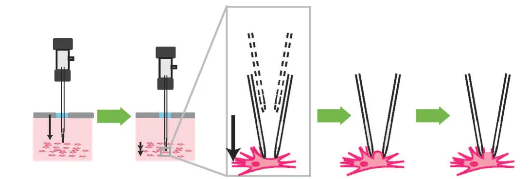
Kodandaramaiah, Boyden and Forest set out to automate a 30-year-old technique known as whole-cell patch clamping, which involves bringing a tiny hollow glass pipette in contact with the cell membrane of a neuron, then opening up a small pore in the membrane to record the electrical activity within the cell. This skill usually takes a graduate student or postdoc several months to learn.
Kodandaramaiah spent about four months learning the manual patch-clamp technique, giving him an appreciation for its difficulty. “When I got reasonably good at it, I could sense that even though it is an art form, it can be reduced to a set of stereotyped tasks and decisions that could be executed by a robot,” he says.
To that end, Kodandaramaiah and his colleagues built a robotic arm that lowers a glass pipette into the brain of an anesthetized mouse with micrometer accuracy. As it moves, the pipette monitors a property called electrical impedance — a measure of how difficult it is for electricity to flow out of the pipette. If there are no cells around, electricity flows and impedance is low. When the tip hits a cell, electricity can’t flow as well and impedance goes up.
The pipette takes two-micrometer steps, measuring impedance 10 times per second. Once it detects a cell, it can stop instantly, preventing it from poking through the membrane. “This is something a robot can do that a human can’t,” Boyden says.
Once the pipette finds a cell, it applies suction to form a seal with the cell’s membrane. Then, the electrode can break through the membrane to record the cell’s internal electrical activity. The robotic system can detect cells with 90 percent accuracy, and establish a connection with the detected cells about 40 percent of the time.
The researchers also showed that their method can be used to determine the shape of the cell by injecting a dye; they are now working on extracting a cell’s contents to read its genetic profile.
Development of the new technology was funded primarily by the National Institutes of Health, the National Science Foundation and the MIT Media Lab.
New era for robotics
The researchers recently created a startup company, Neuromatic Devices, to commercialize the device.
The researchers are now working on scaling up the number of electrodes so they can record from multiple neurons at a time, potentially allowing them to determine how different parts of the brain are connected.
They are also working with collaborators to start classifying the thousands of types of neurons found in the brain. This “parts list” for the brain would identify neurons not only by their shape — which is the most common means of classification — but also by their electrical activity and genetic profile.
“If you really want to know what a neuron is, you can look at the shape, and you can look at how it fires. Then, if you pull out the genetic information, you can really know what’s going on,” Forest says. “Now you know everything. That’s the whole picture.”
Boyden says he believes this is just the beginning of using robotics in neuroscience to study living animals. A robot like this could potentially be used to infuse drugs at targeted points in the brain, or to deliver gene therapy vectors. He hopes it will also inspire neuroscientists to pursue other kinds of robotic automation — such as in optogenetics, the use of light to perturb targeted neural circuits and determine the causal role that neurons play in brain functions.
Neuroscience is one of the few areas of biology in which robots have yet to make a big impact, Boyden says. “The genome project was done by humans and a giant set of robots that would do all the genome sequencing. In directed evolution or in synthetic biology, robots do a lot of the molecular biology,” he says. “In other parts of biology, robots are essential.”
Other co-authors include MIT grad student Giovanni Talei Franzesi and MIT postdoc Brian Y. Chow.
Research News & Publications Office
Georgia Institute of Technology
75 Fifth Street, N.W., Suite 314
Atlanta, Georgia 30308 USA
Media Relations Contacts: Abby Robinson (abby@innovate.gatech.edu; 404-385-3364) or Caroline McCall (cmccall5@mit.edu; 617-253-1682)
Writer: Anne Trafton, MIT News
News Contact
Abby Robinson
Research News and Publications
abby@innovate.gatech.edu
404-385-3364
Apr. 24, 2012
Fibronectin plays a major role in wound healing and embryonic development. The protein, which is located in the extracellular matrix of cells, has also been linked to pathological conditions including cancer and fibrosis.
During physiological processes, fibronectin fibers are believed to experience mechanical forces that strain the fibers and cause dramatic structural modifications that change their biological activity. While understanding the role of fibronectin strain events in development and disease progression is becoming increasingly important, detecting and interrogating these events is difficult.
In a new study, researchers identified molecular probes capable of selectively attaching to fibronectin fibers under different strain states, enabling the detection and examination of fibronectin strain events in both culture and living tissues.
“The mechano-sensitive molecular probes we identified allow us to dynamically examine the relevance of mechanical strain events within the natural cellular microenvironment and correlate these events with specific alterations in fibronectin associated with the progression of disease,” said Thomas Barker, an assistant professor in the Wallace H. Coulter Department of Biomedical Engineering at Georgia Tech and Emory University.
The study was published on April 23, 2012 in the online early edition of the journal Proceedings of the National Academy of Sciences. Barker worked on the study with Georgia Tech graduate student Lizhi Cao and Harry Bermudez, an assistant professor in the University of Massachusetts Amherst Department of Polymer Science and Engineering. The research was supported by the National Institutes of Health.
Researchers have hypothesized that mechanical forces emanating from cells may partially unfold fibronectin and regulate what proteins bind to it. While simulation and tissue culture experiments support this hypothesis, direct evidence that such molecular events occur in living organisms has not yet been presented, according to Barker.
A technique called intramolecular fluorescence resonance energy transfer (FRET) has been used to detect molecular strain events in fibronectin fibers, but the technique has limitations because it cannot be used on living tissues and requires the fibronectin to be chemically labeled.
“The molecular probes we identified can be used to map molecular strain events in native extracellular matrix and living lung tissues,” explained Barker. “The probes can also be used to study the mechanism by which cells control the mechanical forces that alter fibronectin’s conformation, control the exposure of its binding sites and regulate cell signaling.”
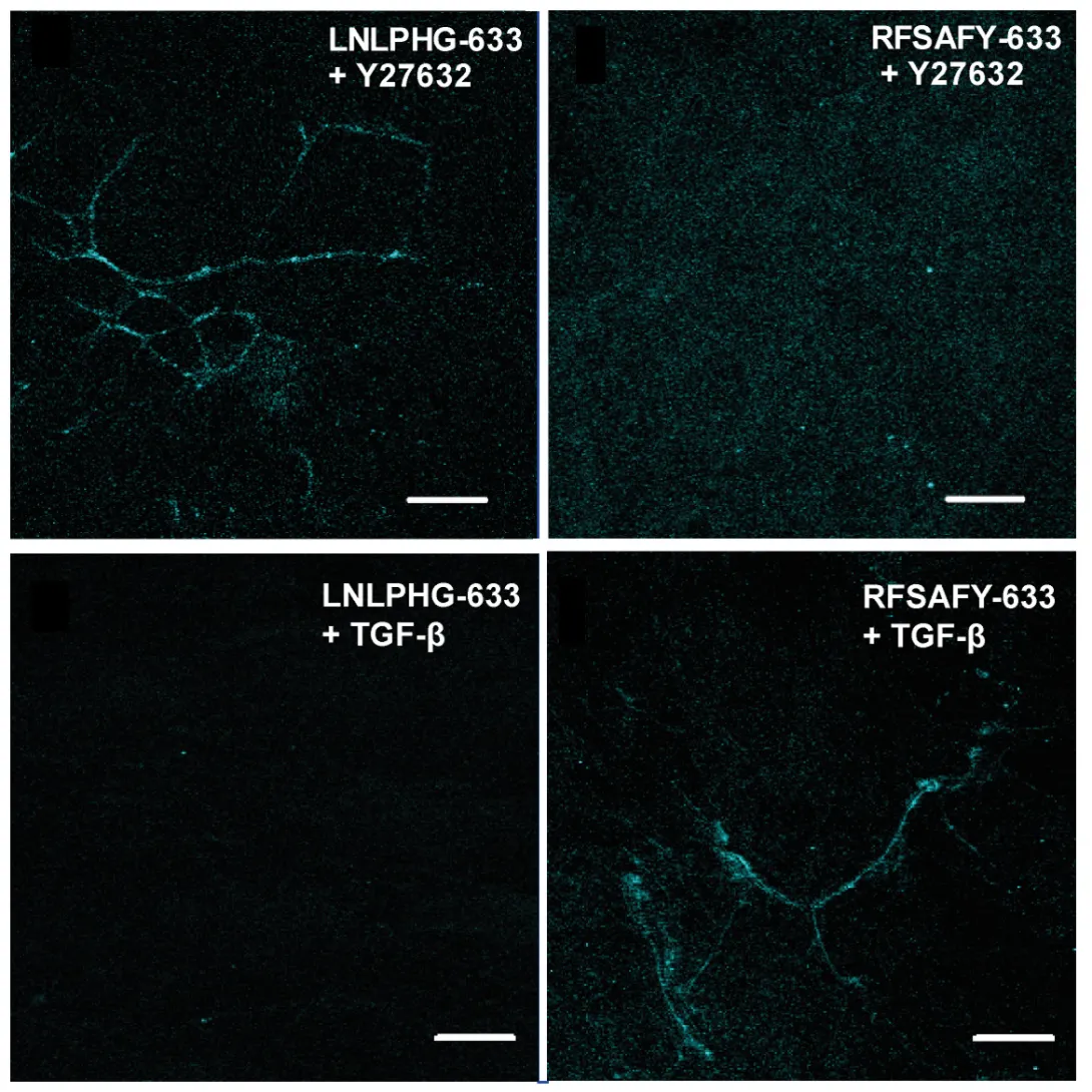
The researchers used a controlled fibronectin fiber deposition and extension technique to apply tension to the fibers and stretch them to 2.6 times their original length without significant breakage. Then they used a technique called phage display to identify peptides capable of discriminating fibronectin fibers under relaxed and strained conditions. The molecular probes displaying peptide sequences LNLPHG and RFSAFY showed the greatest binding affinity to fibronectin fibers and the greatest efficiency in discriminating between relaxed and strained fibers.
For proof-of-concept demonstrations, the researchers used the probes to discriminate fibronectin fibers within native extracellular matrix and mouse lung slices. LNLPHG preferentially attached to relaxed fibronectin fibers, whereas RFSAFY bound to strained fibers. The probes never attached to the same fiber, which confirmed their ability to selectively discriminate regions within a fibronectin fiber network.
“This study strongly suggests that fibronectin fibers under strain display markedly different biochemical signatures that can be used for the molecular-level detection of fibronectin fiber strain,” explained Barker. “The data also show the potential for living tissue to be interrogated for mechano-chemical alterations that lead to physiological and pathological progression.”
In the future, the researchers hope to use these fibronectin strain-sensitive probes to target therapeutics to fibronectin fibers based on their mechanical signature.
This work was supported in part by training grants from the National Institutes of Health (NIH) (Award Nos. T32-GM008433 and T32-EB006343). The content is solely the responsibility of the principal investigators and does not necessarily represent the official views of the NIH.
Research News & Publications Office
Georgia Institute of Technology
75 Fifth Street, N.W., Suite 314
Atlanta, Georgia 30308 USA
Media Relations Contacts: Abby Robinson (abby@innovate.gatech.edu; 404-385-3364) or John Toon (jtoon@gatech.edu; 404-894-6986)
Writer: Abby Robinson
News Contact
Abby Robinson
Research News and Publications
abby@innovate.gatech.edu
404-385-3364
Apr. 02, 2012
Splitting hydrogen and oxygen from water using conventional electrolysis techniques requires considerable amounts of electrical energy. But green plants produce oxygen from water efficiently using a catalytic technique powered by sunlight – a process that is part of photosynthesis and so effective that it is the Earth’s major source of oxygen.
If mimicked by artificial systems, this photocatalytic process could provide abundant new supplies of oxygen and, possibly hydrogen, as a by-product of producing electricity. However, despite its importance to the survival of the planet, scientists don’t fully understand the complex process plants use to harness the sun’s energy.
A paper published April 2 in the journal Proceedings of the National Academy of Sciences moves scientists closer to that understanding by showing the importance of a hydrogen bonding water network in that portion of the photosynthetic machinery known as photosystem II. Using Fourier transform infrared spectroscopy (FT-IR) on photosystem II extracted from ordinary spinach, researchers at the Georgia Institute of Technology tested the idea that a network of hydrogen-bonded water molecules plays a catalytic role in the process that produces oxygen.
“By substituting ammonia, an analog of the water molecule that has a similar structure, we were able to show that the network of hydrogen-bonded water molecules is important to the catalytic process,” said Bridgette Barry, a professor in Georgia Tech’s School of Chemistry and Biochemistry and the Petit Institute for Bioengineering and Biosciences. “Substituting ammonia for water inhibited the activity of the photosystem and disrupted the network. The network could be reestablished by addition of a simple sugar, trehalose.”
The research was supported by the National Science Foundation (NSF) and published in the Early Edition of the journal.
In the chloroplasts of green plants, algae and cyanobacteria, oxygen is produced by the accumulation of photo-induced oxidizing equivalents in a structure known as the oxygen-evolving complex (OEC). The OEC contains manganese and calcium ions. Illumination causes oxidation of manganese ions in the OEC. Short laser flashes can be used to step through the reaction cycle, which involves four sequential light-induced oxidation reactions. Oxygen is produced on the fourth step, and then is released from the OEC.
This so-called S state cycle resets with the binding of the substrate, water. Scientists have proposed that a hydrogen bond network, which includes multiple water molecules bound to manganese ions, calcium ions, and protein amide carbonyl (C=O) groups, forms an electrostatic network surrounding the OEC. In this scenario, the extensive hydrogen-bond network would then serve as a component of the catalyst, which splits off oxygen.
To study the process, Barry and graduate student Brandon Polander used precision FT-IR spectroscopy to describe how the network reacts to a short laser flash. The second harmonic of a pulsed Nd-Yag laser was used as the light source. This illumination causes the OEC to undergo one step in its catalytic cycle, the so-called S1 to S2 transition. An infrared spectrum was recorded before and after a laser flash to the photosystem sample, which was isolated from supermarket spinach.
The exquisite sensitivity of FT-IR spectroscopy allowed them to measure changes in the bond strength of the protein C=O groups. The energies of these C=O groups were used as markers of hydrogen bond strength. The brief laser flash oxidized a manganese ion and caused a change in the strength of the C=O bond, which reported an increase in hydrogen bonding to water molecules. When ammonia was added as an inhibitor, a decrease in C=O hydrogen bonding was observed instead. Addition of trehalose, which is known to change the ordering of water molecules at the surface of proteins, blocked this effect of ammonia.
The study describes the coordinated changes that must occur in the protein to facilitate the reaction and shows that the strength of the hydrogen-bonded network is important.
“This research helps to clarify how ammonia inhibits the photosystem, which is something that researchers have been wondering about for many years,” Barry explained. “Our work suggests that ammonia can inhibit the reaction by disrupting this network of hydrogen bonds.”
The research also suggests that in design of artificial devices that carry out this reaction, sustaining a similar hydrogen-bonding network may be important. The stabilizing effect of trehalose discovered by Polander and Barry may also be important.
Beyond the importance of understanding the photosynthetic process, the work could lead to new techniques for producing hydrogen and oxygen using sunlight. One possibility would be to add a biomimetic photocatalytic process to a photovoltaic system producing electricity from the sun.
“In terms of providing new sources of energy, we still have lessons to learn from plants about how they carry out these critical processes,” Barry said. “It would be a great advance for the planet to have new, sustainable, and inexpensive processes to carry out this reaction.”
Ultimately, she hopes the full water oxidizing cycle can be explored and potentially harnessed or imitated for oxygen and energy production.
“We are only looking at a single part of the overall reaction now, but we would like to study the entire cycle, in which oxygen is produced, to see how the interactions in the water network change and how the interactions with the protein change,” Barry said. “The work is another step in understanding how plants carry out this amazing series of photosynthetic reactions.”
Research News & Publications Office
Georgia Institute of Technology
75 Fifth Street, N.W., Suite 314
Atlanta, Georgia 30308 USA
Media Relations Assistance: John Toon (404-894-6986)(jtoon@gatech.edu) or Abby Robinson (404-385-3364)(abby@innovate.gatech.edu).
Writer: John Toon
News Contact
John Toon
Research News & Publications Office
404-894-6986
jtoon@gatech.edu
Mar. 28, 2012

Treating invasive brain tumors with a combination of chemotherapy and radiation has improved clinical outcomes, but few patients survive longer than two years after diagnosis. The effectiveness of the treatment is limited by the tumor’s aggressive invasion of healthy brain tissue, which restricts chemotherapy access to the cancer cells and complicates surgical removal of the tumor.
To address this challenge, researchers from the Georgia Institute of Technology and Emory University have designed a new treatment approach that appears to halt the spread of cancer cells into normal brain tissue in animal models. The researchers treated animals possessing an invasive tumor with a vesicle carrying a molecule called imipramine blue, followed by conventional doxorubicin chemotherapy. The tumors ceased their invasion of healthy tissue and the animals survived longer than animals treated with chemotherapy alone.
“Our results show that imipramine blue stops tumor invasion into healthy tissue and enhances the efficacy of chemotherapy, which suggests that chemotherapy may be more effective when the target is stationary,” said Ravi Bellamkonda, a professor in the Wallace H. Coulter Department of Biomedical Engineering at Georgia Tech and Emory University. “These results reveal a new strategy for treating brain cancer that could improve clinical outcomes.”
The results of this work were published on March 28, 2012 in the journal Science Translational Medicine. The research was supported primarily by the Ian’s Friends Foundation and partially by the Georgia Cancer Coalition, the Wallace H. Coulter Foundation and a National Science Foundation graduate research fellowship.
In addition to Bellamkonda, collaborators on the project include Jack Arbiser, a professor in the Emory University Department of Dermatology; Daniel Brat, a professor in the Emory University Department of Pathology and Laboratory Medicine; and the paper’s lead author, Jennifer Munson, a former Fulbright Scholar who was a bioengineering graduate student in the Georgia Tech School of Chemical & Biomolecular Engineering when the research was conducted.
Arbiser designed the novel imipramine blue compound, which is an organic triphenylmethane dye. After in vitro experiments showed that imipramine blue effectively inhibited movement of several cancer cell lines, the researchers tested the compound in an animal model of aggressive cancer that exhibited attributes similar to a human brain tumor called glioblastoma.
“There were many reasons why we chose to use the RT2 astrocytoma rat model for these experiments,” said Brat. “The tumor exhibited properties of aggressive growth, invasiveness, angiogenesis and necrosis that are similar to human glioblastoma; the model utilized an intact immune system, which is seen in the human disease; and the model enabled increased visualization by MRI because it was a rat model, rather than a mouse.”
Because imipramine blue is hydrophobic and doxorubicin is cytotoxic, the researchers encapsulated each compound in an artificially-prepared vesicle called a liposome so that the drugs would reach the brain. The liposomal drug delivery vehicle also ensured that the drugs would not be released into tissue until they passed through leaky blood vessel walls, which are only present where a tumor is growing.
Animals received one of the following four treatments: liposomes filled with saline, liposomes filled with imipramine blue, liposomes filled with doxorubicin chemotherapy, or liposomes filled with imipramine blue followed by liposomes filled with doxorubicin chemotherapy.
All of the animals that received the sequential treatment of imipramine blue followed by doxorubicin chemotherapy survived for 200 days -- more than 6 months -- with no observable tumor mass. Of the animals treated with doxorubicin chemotherapy alone, 33 percent were alive after 200 days with a median survival time of 44 days. Animals that received capsules filled with saline or imipramine blue – but no chemotherapy -- did not survive more than 19 days.
“Our results show that the increased effectiveness of the chemotherapy treatment is not because of a synergistic toxicity between imipramine blue and doxorubicin. Imipramine blue is not making the doxorubicin more toxic, it’s simply stopping the movement of the cancer cells and containing the cancer so that the chemotherapy can do a better job,” explained Bellamkonda, who is also the Carol Ann and David D. Flanagan Chair in Biomedical Engineering and a Georgia Cancer Coalition Distinguished Cancer Scholar.
MRI results showed a reduction and compaction of the tumor in animals treated with imipramine blue followed by doxorubicin chemotherapy, while animals treated with chemotherapy alone presented with abnormal tissue and glioma cells. MRI also indicated that the blood-brain barrier breach often seen during tumor growth was present in the animals treated with chemotherapy alone, but not the group treated with chemotherapy and imipramine blue.
According to the researchers, imipramine blue appears to improve the outcome of brain cancer treatment by altering the regulation of actin, a protein found in all eukaryotic cells. Actin mediates a variety of essential biological functions, including the production of reactive oxygen species. Most cancer cells exhibit overproduction of reactive oxygen species, which are thought to stimulate cancer cells to invade healthy tissue. The dye’s reorganization of the actin cytoskeleton is thought to inhibit production of enzymes that produce reactive oxygen species.
“I formulated the imipramine blue compound as a triphenylmethane dye because I knew that another triphenylmethane dye, gentian violet, exhibited anti-cancer properties, and I decided to use imipramine -- a drug used to treat depression -- as the starting material because I knew it could get into the brain,” said Arbiser.
For future studies, the researchers are planning to test imipramine blue’s effect on animal models with invasive brain tumors, metastatic tumors, and other types of cancer such as prostate and breast.
“While we need to conduct future studies to determine if the effect of imipramine blue is the same for different types of cancer diagnosed at different stages, this initial study shows the possibility that imipramine blue may be useful as soon as any tumor is diagnosed, before anti-cancer treatment begins, to create a more treatable tumor and enhance clinical outcome,” noted Bellamkonda.
Research News & Publications Office
Georgia Institute of Technology
75 Fifth Street, N.W., Suite 314
Atlanta, Georgia 30308 USA
Media Relations Contacts: Abby Robinson (abby@innovate.gatech.edu; 404-385-3364) or John Toon (jtoon@gatech.edu; 404-894-6986)
Writer: Abby Robinson
News Contact
Abby Robinson
Research News and Publications
abby@innovate.gatech.edu
404-385-3364