Apr. 04, 2011
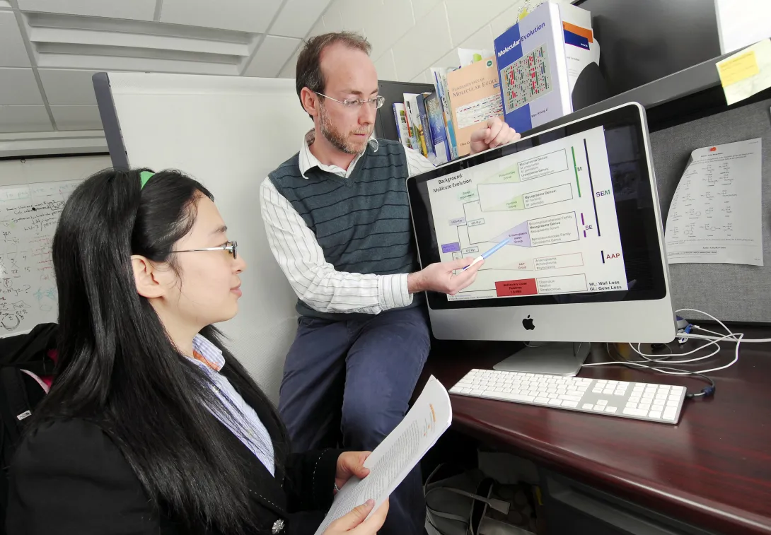
A new study reveals that a group of ancient enzymes adapted to substantial changes in ocean temperature and acidity during the last four billion years, providing evidence that life on Early Earth evolved from a much hotter, more acidic environment to the cooler, less acidic global environment that exists today.
The study found that a group of ancient enzymes known as thioredoxin were chemically stable at temperatures up to 32 degrees Celsius (58 degrees Fahrenheit) higher than their modern counterparts. The enzymes, which were several billion years old, also showed increased activity at lower pH levels -- which correspond to greater acidity.
"This study shows that a group of ubiquitous proteins operated in a hot, acidic environment during early life, which supports the view that the environment progressively cooled and became more alkaline between four billion and 500 million years ago," said Eric Gaucher, an associate professor in the School of Biology at the Georgia Institute of Technology.
The study, which was published April 3 in the advance online edition of the journal Nature Structural & Molecular Biology, was conducted by an international team of researchers from Georgia Tech, Columbia University and the Universidad de Granada in Spain.
Major funding for this study was provided by two grants from the National Aeronautics and Space Administration to Georgia Tech, a grant from the National Institutes of Health to Columbia University, and a grant from the Spanish Ministry of Science and Innovation to the Universidad de Granada.
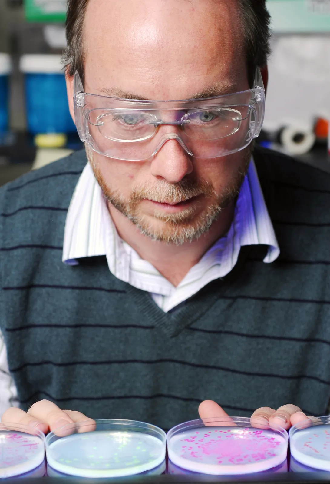
Using a technique called ancestral sequence reconstruction, Gaucher and Georgia Tech biology graduate student Zi-Ming Zhao reconstructed seven ancient thioredoxin enzymes from the three domains of life -- archaea, bacteria and eukaryote -- that date back between one and four billion years.
To resurrect these enzymes, which are found in nearly all known modern organisms and are essential for life in mammals, the researchers first constructed a family tree of the more than 200 thioredoxin sequences available from the three domains of life. Then they reconstructed the sequences of the ancestral thioredoxin enzymes using statistical methods based on maximum likelihood. Finally, they synthesized the genes that encoded these sequences, expressed the ancient proteins in the cells of modern Escherichia coli bacteria and then purified the proteins.
"By resurrecting proteins, we are able to gather valuable information about the adaptation of extinct forms of life to climatic, ecological and physiological alterations that cannot be uncovered through fossil record examinations," said Gaucher.
The reconstructed enzymes from the Precambrian period -- which ended about 542 million years ago -- were used to examine how environmental conditions, including pH and temperature, affected the evolution of the enzymes and their chemical mechanisms.
"Given the ancient origin of the reconstructed thioredoxin enzymes, with some of them predating the buildup of atmospheric oxygen, we thought their catalytic chemistry would be simple, but we found that thioredoxin enzymes use a complex mixture of chemical mechanisms that increases their efficiency over the simpler compounds that were available in early geochemistry," said Julio Fernández, a professor in the Department of Biological Sciences professor at Columbia University.
Fernández led a team that included Columbia University postdoctoral researchers Raul Perez-Jimenez, Jorge Alegre-Cebollada and Sergi Garcia-Manyes, and graduate student Pallav Kosuri in using an assay based on single molecule force spectroscopy to measure the activity level of the thioredoxin enzymes under different pH levels.
For their experiments, the researchers used an atomic force microscope to pick up and stretch an engineered protein in a solution containing thioredoxin. They first applied a constant force to the protein, causing it to rapidly unfold and expose its disulfide bonds to the thioredoxin enzymes. The rate at which a thioredoxin enzyme snipped the disulfide bonds determined the enzyme's level of efficiency.
The study results showed that the three oldest thioredoxin enzymes -- those thought to have inhabited Earth 4.2 to 3.5 billion years ago -- were able to operate in lower pH environments than the modern thioredoxin enzymes.
"Our analysis indicates that ancient thioredoxin enzymes were well adapted to function under acidic conditions and that they maintained their high level of activity as they evolved in more alkaline environments," said Fernández.
To measure the temperature range in which the enzymes operated, professor Jose Sanchez-Ruiz and graduate student Alvaro Inglés-Prieto from the Departamento de Química-Física at the Universidad de Granada in Spain used a technique called differential scanning calorimetry. This method measures the stability of enzymes by heating the enzymes at a constant rate and measuring the heat change associated with their unfolding.
The researchers found that the ancient proteins were stable at temperatures up to 32 degrees Celsius higher than the modern thioredoxins. The experiments showed that the enzymes exhibited higher temperature stability the older they were. The results provide evidence that ancestral thioredoxins adapted to the cooling trend of ancient oceans, as inferred from geological records.
"Our results confirm that life has the remarkable ability to adapt to a wide range of historical environmental conditions; and by extension, life will undoubtedly adapt to future environmental changes, albeit at some cost to many species," said Gaucher.
This study also showed that the experimental resurrection of ancient proteins together with the sensitivity of single-molecule techniques can be a powerful tool for understanding the origin and evolution of life on Earth.
The researchers are currently using this strategy to assess other enzymes to get a clearer picture of what life was like on Early Earth. They are also applying these tools to the field of biotechnology, where enzymes play important roles in many industrial processes.
"The functions and characteristics we observed in the ancestral enzymes show that our techniques can be implemented to generate improved enzymes for a wide range of applications," added Perez-Jimenez.
This project was supported by the National Aeronautics and Space Administration (NASA) (Award Nos. NNX08AO12G and NNA09DA78A). The content is solely the responsibility of the principal investigator and does not necessarily represent the official view of NASA.
Research News & Publications Office
Georgia Institute of Technology
75 Fifth Street, N.W., Suite 314
Atlanta, Georgia 30308 USA
Media Relations Contacts: Abby Robinson (abby@innovate.gatech.edu; 404-385-3364) or John Toon (jtoon@gatech.edu; 404-894-6986)
Writer: Abby Robinson
News Contact
Abby Robinson
Research News and Publications
Contact Abby Robinson
404-385-3364
Mar. 23, 2011

Avian embryos could join the list of model organisms used to study a specific type of cell migration called epiboly, thanks to the results of a study published this month in the journal Developmental Dynamics. The new study provides insights into the mechanisms of epiboly, a developmental process involving mass movement of cells as a sheet, which is linked with medical conditions that include wound healing and cancer.
The study, published online on March 15, explains how epithelial cells expand as a sheet and migrate to engulf the entire avian egg yolk as it grows. It also reveals the presence of certain molecules during this process that have not been previously reported in other major developmental models, including Xenopus frogs and zebrafish.
"These molecules and mechanisms of early development in the avian embryo may demonstrate evolutionary differences across species in the collective movement of epithelial cells and motivate additional studies of avian embryo development," said Evan Zamir, an assistant professor in the George W. Woodruff School of Mechanical Engineering at Georgia Tech.
Matt Futterman, who worked on the project as a graduate student at Georgia Tech, and mechanical engineering professor Andrés García also contributed to this study. The research was funded by Zamir's new faculty support from Georgia Tech and by a grant to García from the National Institutes of Health.
In the study, the researchers conducted immunofluorescence and high-resolution confocal microscopy experiments to examine the spatial distribution and expression of five proteins -- vimentin, cytokeratin, β-catenin, E-cadherin and laminin -- as cells moved to wrap the yolk sac of quail embryos during development.
The results showed that during this process, four of the proteins -- vimentin, cytokeratin, β-catenin and E-cadherin -- appeared in the cells located at the free edge of the migrating cell sheet. Finding dense interconnected networks of both vimentin and cytokeratin in the edge cells surprised the researchers.
"Since cytokeratin is generally associated with the epithelial phenotype and vimentin is generally associated with the mesenchymal phenotype, it's rare to see them expressed in the same cells, but this does occur in metastasizing tumor cells," said Zamir.
Cells expressing the mesenchymal phenotype are typically found in connective tissues -- such as bone, cartilage, and the lymphatic and circulatory systems -- whereas cells of the epithelial phenotype are found in cavities and glands and on surfaces throughout the body.
This finding provides evidence that epithelial cells normally attached to a membrane surface underwent biochemical changes that enabled them to assume a mesenchymal cell phenotype, which enhanced their migratory capacity. This process, called partial epithelial-to-mesenchymal transition, has many similarities to the initiation of tumor cell metastasis and wound healing.
Since this epithelial and mesenchymal expression pattern in the edge cells has not previously been reported in Xenopus or zebrafish, it may be unique to the avian embryo. This discovery would make the avian embryo a valuable model for studying tumor cell migration and wound healing.
In addition to detailing protein expression in the quail embryo during development, the researchers also determined the origin of the new cells required at the migrating edge to cover the growing yolk. During development, the radius of the quail yolk doubles every day for the first few days, representing a hundreds-fold increase in the egg yolk surface area.
"For each individual cell that has to cover the egg yolk as it grows, the migration around the yolk is extraordinary, because it's such a large territory -- it would be like an ant walking across the earth," explained Zamir.
Looking more closely at the edge cells, the researchers found strong evidence that expansion of the edge cell population was due exclusively to cells relocating from an interior region to the edge as the embryo expanded. The cells located at the free edge generated the bulk of the traction force necessary for expansion and towed the cells within the interior of the epithelium.
"These experiments confirm that edge cell proliferation is not the primary mechanism for expansion of the edge cell population," noted Zamir. "And our observation of epithelial-to-mesenchymal transition in the edge cells explains how these epithelial cells might be changing phenotype to become migratory in this rapidly expanding sheet."
To determine if this study's findings are indeed unique to the avian embryo, Zamir plans to conduct further studies to characterize protein expression and cell migration in Xenopus and zebrafish.
Research News & Publications Office
Georgia Institute of Technology
75 Fifth Street, N.W., Suite 314
Atlanta, Georgia 30308 USA
Media Relations Contacts: Abby Robinson (abby@innovate.gatech.edu; 404-385-3364) or John Toon (jtoon@gatech.edu; 404-894-6986)
Writer: Abby Robinson
News Contact
Abby Robinson
Research News and Publications
Contact Abby Robinson
404-385-3364
Mar. 02, 2011
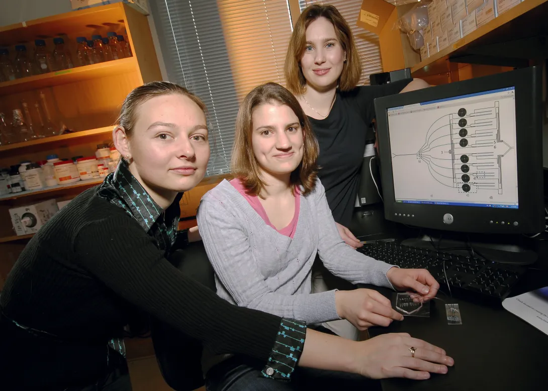
Manipulation of cells by a new microfluidic device may help clinicians improve a promising cancer therapy that harnesses the body's own immune cells to fight such diseases as metastatic melanoma, non-Hodgkin's lymphoma, chronic lymphocytic leukemia and neuroblastoma.
The therapy, known as adoptive T cell transfer, has shown encouraging results in clinical trials. This treatment involves removing disease-fighting immune cells called T cells from a cancer patient, multiplying them in the laboratory and then infusing them back into the patient's body to attack the cancer. The effectiveness of this therapy, however, is limited by the finite lifespan of T cells -- after many divisions, these cells become unresponsive and inactive.
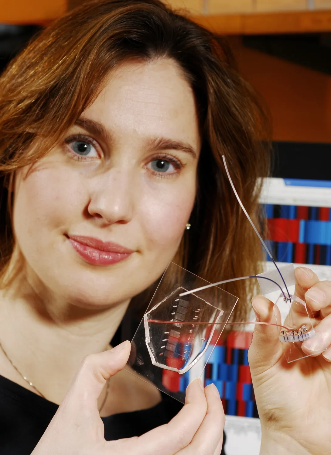
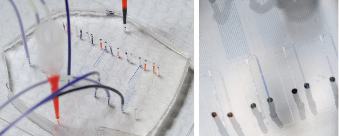
Researchers at Georgia Tech and Emory University have addressed this limitation by developing a microfluidic device for sample handling that allows a statistical model to be generated to evaluate cell responsiveness and accurately predict cell "age" and quality. Being able to assess the age and responsiveness of T cells -- and therefore transfer only young functional cells back into a cancer patient's body -- offers the potential to improve the therapeutic outcome of several cancers.
"The statistical model, enabled by the data generated with the microfluidic device, revealed an optimal combination of extracellular and intracellular proteins that accurately predict T cell age," said Melissa Kemp, an assistant professor in the Wallace H. Coulter Department of Biomedical Engineering at Georgia Tech and Emory University. "Knowing this information will help facilitate the clinical development of appropriate T cell expansion and selection protocols."
Details on the microfluidic device and statistical model were published in the March issue of the journal Molecular & Cellular Proteomics. This work was supported by the National Institutes of Health, Georgia Cancer Coalition, and Georgia Tech Integrative Biosystems Institute.
Currently, clinicians measure T cell age by using multiple assays that rely on measurements from large cell populations. The measurements determine if cells are exhibiting functions known to appear at different stages in the life cycle of a T cell.
"Since no one measurement is a perfect predictor, it is advantageous to concurrently sample multiple proteins at different time points, which we can do with our microfluidic device," explained Kemp, who is also a Georgia Cancer Coalition Distinguished Professor. "The wealth of information we get from our device for a small number of cells far exceeds a single measurement from a population the same size by another assay type."
For their study, Kemp, electrical engineering graduate student Catherine Rivet and biomedical engineering undergraduate student Abby Hill analyzed CD8+ T cells from healthy blood donors. They acquired information from 25 static biomarkers and 48 dynamic signaling measurements and found a combination of phenotypic markers and protein signaling dynamics -- including Lck, ERK, CD28 and CD27 -- to be the most useful in predicting cellular age.
To obtain biomarker and dynamic signaling event measurements, the researchers ran the donor T cells through a microfluidic device designed in collaboration with Hang Lu, an associate professor in the Georgia Tech School of Chemical & Biomolecular Engineering. After stimulating the cells, the device divided them into different channels corresponding to eight different time points, ranging from 30 seconds to seven minutes. Then they were divided again into populations that were chemically treated to halt the biochemical reactions at snapshots in time to build up a picture of the signaling events that occurred as the T cells responded to antigen.
"While donor-to-donor variability is a confounding factor in these types of experiments, the technological platform minimized the experimental data variance and allowed stimulation time to be precisely controlled," said Lu.
With the donor T cell data, the researchers developed a model to assess which biomarkers or dynamical signaling events best predicted the quality of T cell function. The model found the most informative data in predicting cellular age to be the initial changes in signaling dynamics.
"Although a combination of biomarker and dynamic signaling data provided the optimal model, our results suggest that signaling information alone can predict cellular age almost as well as the entire dataset," noted Kemp.
In the future, Kemp plans to use this approach of combining multiple cell-based experiments on a microfluidic chip to integrate single-cell information with population-averaged techniques, such as multiplexed immunoassays or mass spectrometry.
This project is supported in part by the National Institutes of Health (NIH)(Grant No. R21CA134299). The content is solely the responsibility of the principal investigator and does not necessarily represent the official views of the NIH.
Research News & Publications Office
Georgia Institute of Technology
75 Fifth Street, N.W., Suite 314
Atlanta, Georgia 30308 USA
Media Relations Contacts: Abby Robinson (abby@innovate.gatech.edu; 404-385-3364) or John Toon (jtoon@gatech.edu; 404-894-6986)
Writer: Abby Robinson
News Contact
Abby Robinson
Research News and Publications
Contact Abby Robinson
404-385-3364
Feb. 10, 2011
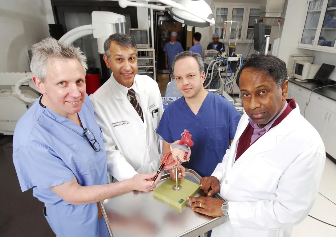
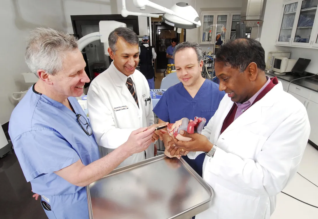
A Georgia Tech and Emory University medical device startup that has developed a system to simplify and standardize the technique for opening and closing the beating heart during cardiac surgery has received a $5.1 million investment.
Apica Cardiovascular has licensed the Georgia Tech/Emory technology and will further develop the system, which will make the transapical access and closure procedure required for delivering therapeutic devices to the heart more routine for all surgeons. The goal is to expand the use of surgery techniques that are less invasive and do not require stopping the heart.
"Our company has leveraged the expertise in cardiovascular technology at Georgia Tech and the clinical experience of surgeons at Emory University to develop a technology that has the potential to revolutionize the delivery of different types of medical devices to the heart, including aortic and mitral valves," said the company's CEO James Greene.
With research and development support from the Coulter Foundation Translational Research Program and the Georgia Research Alliance VentureLab program, the company has already completed a series of pre-clinical studies to test the functionality of the device and its biocompatibility.
The improved heart surgery system consists of a conduit with proprietary technology inside that allows the conduit to be securely attached to the beating heart. Surgeons can then deliver therapeutic devices, such as heart valves or left ventricular assist devices, into the beating heart without loss of blood or exposure to air. Once a therapeutic device has been delivered and surgery is complete, the company's system closes and seals the access site with a biocompatible implant. The closure site can be reopened if necessary.
"By minimizing the incision size to gain access to the beating heart and eliminating the need for conventional sutures, our system improves safety, decreases procedure time and reduces the technical challenges associated with these new minimally invasive procedures," explained Vinod Thourani, an associate professor of surgery and associate director of the Structural Heart Center in Emory University's Division of Cardiothoracic Surgery.
With the new investment from Ireland-based Seroba Kernel Life Sciences and Israel-based TriVentures, the company will continue to conduct research and pre-clinical trials in Atlanta, ultimately leading up to regulatory approval. These efforts will be led by Jorge H. Jimenez, the chief technology officer of the company, which is in the VentureLab process at ATDC, Georgia Tech’s startup company accelerator.
"Our goal is to accelerate and expand the adoption of less-invasive therapeutic procedures to a greater number of surgeons and as a result, many underserved patients will receive needed treatment for valve disease and end-stage heart failure," said Ajit Yoganathan, Regents professor and Wallace H. Coulter Distinguished Faculty Chair in Biomedical Engineering in the Wallace H. Coulter Department of Biomedical Engineering at Georgia Tech and Emory University.
The startup will also have an office in Ireland, which will benefit from the strong research collaborations between Georgia Tech, Georgia Tech Ireland and the National University of Ireland, Galway.
"We seek to contribute to and benefit from a global innovation ecosystem in ways that accelerate research results to the market while enhancing economic development opportunities here in Georgia," said Stephen E. Cross, Georgia Tech's executive vice president for research. "Apica Cardiovascular is a perfect example of the synergy between our leading edge work in Atlanta, our Irish translational unit GT Ireland, and our partnership with the National University of Ireland, Galway."
Apica Cardiovascular was founded in 2009 based on technology invented by Jimenez, Thourani, Yoganathan and Thomas Vassiliades, who was an associate professor of cardiothoracic surgery at Emory University at the time. The company was named Emory University's Startup Company of 2010.
About ATDC:
The Advanced Technology Development Center (ATDC) is a startup accelerator that helps technology entrepreneurs in Georgia launch and build successful companies. Founded in 1980, ATDC has graduated more than 120 companies, which together have raised more than a billion dollars in outside financing. In 2010, ATDC was named to Forbes Magazine’s list of the “10 technology incubators that are changing the world.”
Research News & Publications Office
Georgia Institute of Technology
75 Fifth Street, N.W., Suite 314
Atlanta, Georgia 30308 USA
Media Relations Contacts: Abby Robinson (abby@innovate.gatech.edu; 404-385-3364) or John Toon (jtoon@gatech.edu; 404-894-6986)
Writer: Abby Robinson
Aug. 10, 2010


The Georgia Institute of Technology has received a EUREKA grant from the National Institutes of Health (NIH) to design a new way to treat invasive brain tumors by capturing the migrating cells that spread the disease. The EUREKA -- Exceptional, Unconventional Research Enabling Knowledge Acceleration -- program helps scientists test new, unconventional ideas or tackle major methodological or technical challenges.
The research team plans to develop a system that will excavate brain tumor cells by directing them away from their location in the interior of the brain to a more external location where they can be removed or killed. Nanofiber-based polymer thin films coated with biochemical cues will be aligned in the brain to provide a corridor for tumor cells to follow to a gel-based ‘sink’ where they will be captured and safely removed or encouraged to die through chemical signaling.
“We believe this is the first attempt to exploit the invasive, migrating properties of brain tumors by engineering a path for the tumors to move away from the primary site to a location where treatment can occur,” said lead investigator Ravi Bellamkonda, a professor in the Wallace H. Coulter Department of Biomedical Engineering at Georgia Tech and Emory University.
Collaborating with Bellamkonda on this project are Tobey MacDonald, director of the pediatric neuro-oncology program at the Aflac Cancer Center and Blood Disorders Service of Children’s Healthcare of Atlanta and an associate professor of pediatrics at the Emory University School of Medicine; and Barun Brahma, a pediatric neurosurgeon at Children’s Healthcare of Atlanta. The initial partnership between the researchers began with seed funding from the Georgia Cancer Coalition and Ian’s Friends Foundation.
The National Cancer Institute is providing more than $1 million for the EUREKA grant. For the project, Bellamkonda, MacDonald and Brahma are focusing on treating medulloblastomas -- highly malignant brain tumors that account for more than 20 percent of pediatric brain tumors.
“Medulloblastoma is the most common malignant brain tumor we see in children, but unfortunately the five-year survival rates for children with this cancer only range from 50 to 70 percent and the majority of survivors have a significantly reduced quality of life as a result of treatment-related toxicities,” said MacDonald, who is also a Georgia Cancer Coalition Distinguished Scholar. “An increasing number of survivors are also at risk for developing secondary malignancies as a result of the treatment we now administer. Clearly we have to do a much better job at treating these tumors; however, improving survival while reducing the toxic effects of treatment will require a highly innovative approach.”
Medulloblastoma treatment currently involves surgery followed by radiation therapy to the entire brain and spine and up to one year of intensive intravenous chemotherapy. However, radiation is often delayed or omitted altogether in young children due to its debilitating long-term side effects on the developing central nervous system.
These changes to the timing of radiation administration can adversely impact survival. And while surgery is a mainstay of treatment, it too can cause a significant loss of cognitive and neurological function due to the critical areas of the brain that may be involved by the tumor’s spread but require an extensive surgical area to remove as much of the tumor as possible.
This EUREKA grant aims to address the urgent need to develop therapies to safely treat invasive medulloblastomas in children.
“Our plan is to deliver the tumor to the drug -- by directing tumor cells to a specially engineered gel that can be removed or designed to kill the cells -- rather than the current strategy of delivering the drug to the tumor, which is problematic due to the irregular vasculature and poor diffusivity of the tumor tissue,” explained Bellamkonda, who is also a Georgia Cancer Coalition Distinguished Scholar.
The researchers plan to design a polymer thin film system that will include topographical and biochemical cues similar to those that guide the initial brain tumor invasion. The thin films will be rolled up and deployed with minimally invasive catheters. Because neural tissue will not be suctioned and the films are very thin, there should be minimal tissue and tumor disruption.
The films will also be non-toxic to the patient because they will be engineered with biocompatible, stable polymers. In previous studies, the polymers have been implanted in the nervous systems of small animals for more than 16 weeks with no adverse tissue reactions.
“This research represents a radical approach to treating invasive tumors that is based on the universal properties and mechanics of cell motility and the migration characteristic of metastasis, regardless of the molecular and genetic origins of the tumor,” added Bellamkonda.
If successful, this approach would identify a new and innovative way to treat pediatric medulloblastomas and has the potential to open a new avenue for the treatment of other invasive solid tumors, such as brain stem tumors. These cancers are incurable because they are located in an inoperable region and/or they are resistant or inaccessible to the delivery of chemotherapy agents.
Research News & Publications Office
Georgia Institute of Technology
75 Fifth Street, N.W., Suite 314
Atlanta, Georgia 30308 USA
Media Relations Contacts: Abby Vogel Robinson (404-385-3364; abby@innovate.gatech.edu) or John Toon (404-894-6986; jtoon@gatech.edu)
Writer: Abby Vogel Robinson
News Contact
Abby Vogel Robinson
Research News and Publications
Contact Abby Vogel Robinson
404-385-3364