Sep. 20, 2011

The National Institutes of Health (NIH) has awarded nearly $2 million to researchers at the Georgia Institute of Technology and Emory University to develop a new class of therapeutics for treating traumatic injuries and degenerative diseases.
The five-year project focuses on developing biomaterials capable of capturing certain molecules from embryonic stem cells and delivering them to wound sites to enhance tissue regeneration in adults. By applying these unique molecules, clinicians may be able to harness the regenerative power of stem cells while avoiding concerns of tumor formation and immune system compatibility associated with most stem cell transplantation approaches.
"Pre-clinical and clinical evidence strongly suggests that the biomolecules produced by stem cells significantly impact tissue regeneration independent of differentiation into functionally competent cells," said Todd McDevitt, director of the Stem Cell Engineering Center at Georgia Tech and an associate professor in the Wallace H. Coulter Department of Biomedical Engineering at Georgia Tech and Emory University. "We want to find out if the signaling molecules responsible for scarless wound healing and functional tissue restoration during early stages of embryological development can be used with adult wounds to produce successful tissue regeneration without scar formation."
In addition to McDevitt, Coulter Department associate professor Johnna Temenoff and Woodruff School of Mechanical Engineering professor Robert Guldberg are also investigators on the project.
Regenerative medicine seeks to restore normal structure and function to tissues compromised by degenerative diseases and traumatic injuries. The contrast between embryonic and adult wound healing suggests that molecules that facilitate tissue regeneration during embryonic development are distinctly different from those of adult tissues.
This grant includes plans for engineering biomaterials that can efficiently capture morphogens, which are molecules secreted by embryonic stem cells undergoing differentiation. The study will also evaluate the regenerative activity of molecule-filled biomaterials in animal models of dermal wound healing, hind limb ischemia and bone fractures. Examining the effects of the morphogens on a range of animal wound models will increase the likelihood of success and define any limitations of the technology, such as its use for specific tissues or injuries.
"Biomaterials have largely been used in an attempt to direct stem cell differentiation or serve as passive cell transplantation vehicles for regenerative medicine and tissue engineering purposes," said McDevitt, who is also a Petit Faculty Fellow in the Institute for Bioengineering and Bioscience at Georgia Tech. "The idea of specifically engineering biomaterial properties to capture and deliver complex assemblies of stem cell-derived morphogens without transplanting the cells themselves represents a novel strategy to translate the potency of stem cells into a viable regenerative medicine therapy."
The award was one of 17 granted this year through the NIH Director's Transformative Research Projects Program (T-R01), which was created to challenge the status quo with innovative ideas that have the potential to advance fields and speed the translation of research into improved health for the American public.
Another T-R01 grant was awarded to Coulter Department professor Shuming Nie, associate professor May Wang and University of Pennsylvania School of Medicine Thoracic Surgery Research Laboratory director Sunil Singhal. That $7 million, five-year grant will support continuing work by the Emory-Georgia Tech Nanotechnology Center for Personalized and Predictive Oncology team on developing fluorescent nanoparticle probes that hone in on cancer cells and on creating instruments that visualize them for cancer detection during surgery.
Since its inception in 2009, the NIH Director's Award Program has funded a total of 406 high-risk research projects, including 79 T-R01 awards.
"The NIH Director's Award programs reinvigorate the biomedical work force by providing unique opportunities to conduct research that is neither incremental nor conventional," said James M. Anderson, director of the Division of Program Coordination, Planning and Strategic Initiatives, who guides the NIH Common Fund's High-Risk Research program. "The awards are intended to catalyze giant leaps forward for any area of biomedical research, allowing investigators to go in entirely new directions."
Research News & Publications Office
Georgia Institute of Technology
75 Fifth Street, N.W., Suite 314
Atlanta, Georgia 30308 USA
Media Relations Contacts: Abby Robinson (abby@innovate.gatech.edu; 404-385-3364) or John Toon (jtoon@gatech.edu; 404-894-6986)
Writer: Abby Robinson
News Contact
Abby Robinson
Research News and Publications
Contact Abby Robinson
404-385-3364
Sep. 06, 2011
Engineers and surgeons are working together to improve the treatment of babies born with craniosynostosis, a condition that causes the bone plates in the skull to fuse too soon. Treating this condition typically requires surgery after birth to remove portions of the fused skull bones, and in some cases the bones grow together again too quickly -- requiring additional surgeries.
Researchers in the Atlanta-based Center for Pediatric Healthcare Technology Innovation are developing imaging techniques designed to predict whether a child's skull bones are likely to grow back together too quickly after surgery. They are also developing technologies that may delay a repeat of the premature fusion process.
"Babies are usually only a few months old during the first operation, which lasts more than three hours and requires a unit of blood and a stay in the intensive care unit, so our goal is to develop technologies that will simplify the initial surgery and limit affected babies to this one operation," said center co-director Joseph Williams, clinical director of craniofacial plastic surgery at Children's Healthcare of Atlanta at Scottish Rite and clinical assistant professor in the Department of Plastic and Reconstructive Surgery at Emory University.
Craniosynostosis affects approximately one in every 2,500 babies in the United States. The condition is caused by the premature closure of sutures with bone. Sutures, which are made of tissue that is more flexible than bone, play an important role in brain growth by providing a method for the skull to increase in size. If the sutures close too soon and get replaced with bony tissue, the skull may limit the normal expansion of the brain.
If untreated, craniosynostosis can cause a range of developmental problems. If treated using the standard treatment course, surgeons remove the fused skull bones, break them up, reposition them, and hold them in place with plates and screws. This usually slows bone growth between the bone pieces, allowing room for expansion of the brain. However, studies show that more than six percent of babies need a second operation to separate the bones again and 25 percent of those require a third operation.
"Following the first surgery, there's a clinical need to be able to screen children on a regular basis to predict when their skull bones are going to fuse together again so that the surgeons can determine if additional intervention will be required," said center director Barbara Boyan, the Price Gilbert, Jr. Chair in Tissue Engineering in the Wallace H. Coulter Department of Biomedical Engineering at Georgia Tech and Emory University and associate dean for research and innovation in the Georgia Tech College of Engineering.
To address this need, the researchers have developed a non-invasive technique to monitor bone growth with computed tomography images. They created software that identifies bone in the images, quantifies the distance between the bones, the mass of bone in the gap, and the area and volume of the gap. The research team has demonstrated the utility of this "snake" algorithm using a mouse model of cranial development and recently presented their findings at the 2011 Plastic Surgery Education Foundation conference.
"Using our snake algorithm to analyze computed tomography images of developing skulls in mice, we were able to monitor different types and speeds of bone growth on a daily basis for many weeks," said Chris Hermann, an M.D./Ph.D. student in the Coulter Department. "While one suture fused between 12 and 20 days and then significantly increased in mass for the next 20 days, another came closer together and increased in mass but remained largely open."
The research team recently adapted the technology for use in children and began a clinical study to determine the effectiveness of the algorithm to diagnose cases of craniosynostosis. The researchers hope this technology will improve the ability of physicians to diagnose and determine the severity of craniosynostosis.
In addition, the researchers are studying the biological basis of the condition and developing technologies they hope will delay bone growth and eliminate the need for additional operations. In one project, Coulter Department research scientist Rene Olivares-Navarrete and Williams are examining individuals with craniosynostosis to identify genes that influence suture fusion. Determining the genes that control suture closure may help the researchers identify potential therapeutic targets to prevent premature suture fusion.
The research team has also designed a gel to be injected into the gap created between skull bones during the first surgery. The material -- called a hydrogel because it contains a significant amount of water -- would deliver specific proteins to the area to delay, but not prevent, bone growth.
"The hydrogel cross-links spontaneously because of a reaction between a polyethylene-glycol monomer and a cross-linking molecule, allowing for polymerization without the use of chemical initiators or the production of free radicals," explained Hermann.
Preliminary results in a mouse model of cranial development indicate that the gel, developed in collaboration with Coulter Department associate professor Niren Murthy, can be injected into a gap between skull bones, firm up rapidly and not injure underlying soft tissues or impair bone healing. These pre-clinical results were presented at the Society for Biomaterials Annual Meeting in April 2011.
Both Boyan and Williams see promise in using these technologies to improve the treatment of children with craniosynostosis and eliminate additional operations sometimes needed to treat the condition.
"During the initial surgery, injecting the gel may reduce the operation's severity if it eliminates the need for plates and screws to hold the skull bones in place afterward," explained Boyan, who is also a Georgia Research Alliance (GRA) Eminent Scholar. "After the surgery, if the computed tomography images tell us that the skull is closing too quickly, we may be able to inject the gel through the skin overlying the skull without surgery to further delay the bones from fusing."
The researchers are currently improving the protein release kinetics of the hydrogel and conducting pre-clinical experiments to determine which proteins successfully delay bone growth when included in the gel. Approval from the Food and Drug Administration will be required before this system and hydrogel can be used as a treatment for craniosynostosis.
The Center for Pediatric Healthcare Technology Innovation is supported by Children's Healthcare of Atlanta, in collaboration with Georgia Tech.
Research News & Publications Office
Georgia Institute of Technology
75 Fifth Street, N.W., Suite 314
Atlanta, Georgia 30308 USA
Media Relations Contacts: Abby Robinson (abby@innovate.gatech.edu; 404-385-3364) or John Toon (jtoon@gatech.edu; 404-894-6986)
Writer: Abby Robinson
News Contact
Abby Robinson
Research News and Publications
Contact Abby Robinson
404-385-3364
Aug. 09, 2011
A new study into the causes of leakage in one of the heart's most complex valve structures could lead to improved diagnosis and treatment of the condition.
An estimated 1.6 million Americans suffer moderate to severe leakage through their tricuspid valve, a complex structure that closes off the heart's right ventricle from the right atrium. Most people have at least some leakage in the valve, but what causes the problem is not well understood.
A new study, published online in the journal Circulation on August 1, 2011, found that either dilating the tricuspid valve opening or displacing the papillary muscles that control its operation can cause the valve to leak. A combination of the two actions can increase the severity of the leakage, which is called tricuspid regurgitation.
"We think this is the first in vitro investigation into the mechanics of the tricuspid valve, and that our findings into the mechanisms that cause tricuspid regurgitation could lead to improved diagnosis and treatment," said Ajit Yoganathan, Regents professor and Wallace H. Coulter Distinguished Faculty Chair in Biomedical Engineering in the Wallace H. Coulter Department of Biomedical Engineering at Georgia Tech and Emory University.
The tricuspid valve consists of three flaps that open to allow blood to flow from the heart's upper right chamber to the ventricle. To close the valve, the flaps re-cover the opening, keeping blood from flowing back into the chamber it just left. When the valve is leaky or doesn't close tightly enough, blood flows backward into the chamber just after the heart contracts.
Tricuspid regurgitation has been increasingly recognized as a source of disease in patients with chronic mitral valve regurgitation, but surgical repair of the tricuspid valve alone is recommended only in rare cases. If an individual suffers from severe tricuspid regurgitation, surgeons will sometimes repair the tricuspid valve during a surgery to repair other leaky heart valves.
"Standard clinical procedures that detail when and how tricuspid valve repairs should be performed need to be developed and this study suggests several items that should be considered," said one of the study’s co-authors David H. Adams, chair of the Department of Cardiothoracic Surgery at Mount Sinai Medical Center in New York City. "Current repairs for tricuspid regurgitation focus mainly on remodeling the valve's opening to correct enlargement, but this study shows that it may also be important to restore the position of the papillary muscles, providing as much overlap as possible, in order to conduct effective and durable tricuspid valve repairs."
With funding from the American Heart Association, Yoganathan and Coulter Department graduate student Erin Spinner conducted experiments with porcine tricuspid valves to determine possible causes of tricuspid regurgitation. The valves were attached to a plate designed to create physiological dilation and then placed inside a right heart simulator.
The researchers first investigated the effect of dilating the opening of the tricuspid valves. When the openings stretched to an area 40 percent larger than their normal size, a hole appeared in the central region of the valve. The hole caused leakage, and increased in size with further dilatation. This finding surprised the researchers because similar studies using the same method had shown that the heart's mitral valve could withstand dilation of 75 percent before leaking.
"These results tell us that the tricuspid valve is a much more complex valve than the mitral valve, which only has two flaps," explained Spinner. "Forming a proper seal over the valve opening might be more difficult with three flaps, which could be why such a large percentage of the population experiences some level of tricuspid regurgitation and why some individuals with annular dilation have tricuspid regurgitation and others do not."
The research team also investigated the effect of displacing the valve's three papillary muscles, which attach to the valve via threads. Contraction of the papillary muscles opens the valve and relaxation of the muscles closes the valve. The study showed that papillary displacement alone resulted in significant tricuspid leakage.
"While isolated displacement of the papillary muscles is rare, these results are relevant toward understanding what may happen if the size of the valve opening is repaired, but the papillary muscles are left displaced," noted Yoganathan.
The study also showed that higher levels of tricuspid leakage resulted when the researchers combined the conditions -- dilation of 40 percent or greater and displacement of all papillary muscles.
In their future work, the Coulter Department researchers plan to look at the effect of pulmonary hypertension on the tricuspid valve, because tricuspid regurgitation usually develops in association with pulmonary hypertension -- which is abnormally high blood pressure in the lungs. They also plan to work with their clinical collaborators to extend their findings to humans.
"In our in vitro study we were able to select specific porcine valves, but with human subjects there will be more anatomical variety. For example, two people may have valves of the same diameter, but one person may have longer flaps that are able to compensate for dilation whereas the other might not," noted Yoganathan.
In addition to those already mentioned, Pedro del Nido, a professor in the Department of Cardiothoracic Surgery at Children's Hospital Boston; Emir Veledar, an assistant professor with a joint appointment in the Division of Cardiology and Division of Epidemiology at Emory University; and Coulter Department research engineer Jorge Jimenez and undergraduate students Patrick Shannon and Dana Buice also contributed to this work.
Research News & Publications Office
Georgia Institute of Technology
75 Fifth Street, N.W., Suite 314
Atlanta, Georgia 30308 USA
Media Relations Contacts: Abby Robinson (abby@innovate.gatech.edu; 404-385-3364) or John Toon (jtoon@gatech.edu; 404-894-6986)
Writer: Abby Robinson
News Contact
Abby Robinson
Research News and Publications
Contact Abby Robinson
404-385-3364
Jul. 18, 2011
A new family of contrast agents that sneak into bacteria disguised as glucose food can detect bacterial infections in animals with high sensitivity and specificity. These agents -- called maltodextrin-based imaging probes -- can also distinguish a bacterial infection from other inflammatory conditions.
"These contrast agents fill the need for probes that can accurately image small numbers of bacteria in vivo and distinguish infections from other pathologies like cancer," said Niren Murthy, an associate professor in the Wallace H. Coulter Department of Biomedical Engineering at Georgia Tech and Emory University. "These probes could ultimately improve the diagnosis and treatment of bacterial infections, which remains a major challenge in medicine."
The imaging probes were described in the July 17, 2011 advance online edition of the journal Nature Materials. The research was sponsored by the National Science Foundation and National Institutes of Health.
Coulter Department postdoctoral fellows Xinghai Ning and Seungjun Lee led the project. University of Georgia Complex Carbohydrate Research Center postdoctoral associate Zhirui Wang; and Georgia State University Department of Biology associate professor Eric Gilbert and student Bryan Subblefield also contributed to the work.
In the United States in 2010, bacterial infections caused 40,000 deaths from sepsis and were the leading cause of limb amputations. A major limitation preventing the effective treatment of bacterial infections is an inability to detect them inside the body with accuracy and sensitivity. To image bacterial infections, probes must first deliver a large quantity of the contrast agent into bacteria.
"Most existing imaging probes target the bacterial cell wall and cannot access the inside of the bacteria, but maltodextrin-based imaging probes target a bacterial ingestion pathway, which allows the contrast agent to reach a high concentration within bacteria," said Murthy.
Maltodextrin-based imaging probes consist of a fluorescent dye linked to maltohexaose, which is a major source of glucose for bacteria. The probes deliver the contrast agent into bacteria through the organism's maltodextrin transporter, which only exists in bacterial cells and not mammalian cells.
"To our knowledge, this represents the first demonstration of a targeting strategy that can deliver millimolar concentrations of an imaging probe within bacteria," noted Murthy.
In experiments using a rat model, the researchers found that the contrast agent accumulated in bacteria-infected tissues, but was efficiently cleared from uninfected tissues. They saw a 42-fold increase in fluorescence intensity between bacterial infected and uninfected tissues. However, the contrast agent did not accumulate in the healthy bacterial microflora located in the intestines. Because systemically administered glucose molecules cannot access the interior of the intestines, the bacteria located there never came into contact with the probe.
They also found that the probes could detect as few as one million viable bacteria cells. Current contrast agents for imaging bacteria require at least 100 million bacteria, according to the researchers.
In another experiment, the researchers found that the maltodextrin-based probes could distinguish between bacterial infections and inflammation with high specificity. Tissues infected with E. coli bacteria exhibited a 17-fold increase in fluorescence intensity when compared with inflamed tissues that were not infected.
Additional laboratory experiments showed that the probes could deliver large quantities of imaging probes to gram-positive and gram-negative bacteria for internalization. Both types of bacteria internalized the maltodextrin-based probes at a rate three orders of magnitude faster than mammalian cells.
"Maltodextrin-based probes show promise for imaging infections in a wide range of tissues, with an ability to detect bacteria in vivo with a sensitivity two orders of magnitude higher than previously reported," said Murthy.
This project is supported by the National Science Foundation (NSF) (NSF Career Award No. BES-0546962) and the National Institutes of Health (NIH) (Award Nos. RO1 HL096796-01 and HHSN268201000043C). The content is solely the responsibility of the principal investigator and does not necessarily represent the official views of the NSF or NIH.
Research News & Publications Office
Georgia Institute of Technology
75 Fifth Street, N.W., Suite 314
Atlanta, Georgia 30308 USA
Media Relations Contacts: Abby Vogel Robinson (abby@innovate.gatech.edu; 404-385-3364) or John Toon (jtoon@gatech.edu; 404-894-6986)
Writer: Abby Vogel Robinson
News Contact
Abby Robinson
Research News and Publications
Contact Abby Robinson
404-385-3364
Jun. 16, 2011
Stem cell technologies have been proposed for cell-based diagnostics and regenerative medicine therapies. However, being able to make stem cells efficiently develop into a desired cell type -- such as muscle, skin, blood vessels, bone or neurons -- limits the clinical potential of these technologies.
New research presented on June 16, 2011 at the annual meeting of the International Society for Stem Cell Research (ISSCR) shows that systematically controlling the local and global environments during stem cell development helps to effectively direct the process of differentiation. In the future, these findings could be used to develop manufacturing procedures for producing large quantities of stem cells for diagnostic and therapeutic applications. The research is sponsored by the National Science Foundation and the National Institutes of Health.
"Stem cells don't make any decisions in isolation; their decisions are spatially and temporally directed by biochemical and mechanical cues in their environment," said Todd McDevitt, director of the Stem Cell Engineering Center at Georgia Tech and an associate professor in the Wallace H. Coulter Department of Biomedical Engineering at Georgia Tech and Emory University. "We have designed systems that allow us to tightly control these properties during stem cell differentiation, but also give us the flexibility to introduce a new growth factor or shake the cells a little faster to see how changes like these affect the outcome."
These systems can also be used to compare the suitability of specific stem cell types for a particular use.
"We have developed several platforms that will allow us to conduct head-to-head studies with different kinds of stem cells to determine if one type of stem cell outperforms another type for a certain application," said McDevitt, who is also a Petit Faculty Fellow in the Institute for Bioengineering and Bioscience at Georgia Tech.
Many laboratory growth methods allow stem cells to aggregate in three-dimensional clumps called "embryoid bodies" during differentiation. McDevitt and biomedical engineering graduate student Andres Bratt-Leal incorporated biomaterial particles directly within these aggregates during their formation. They introduced microparticles made of gelatin, poly(lactic-co-glycolic acid) (PLGA) or agarose and tested their impact on the assembly, intercellular communication and morphogenesis of the stem cell aggregates under different conditions by varying the microsphere-to-cell ratio and the size of the microspheres.
The researchers found that the presence of the biomaterials alone modulated embryoid body differentiation, but did not adversely affect cell viability. Compared to typical delivery methods, providing differentiation factors -- retinoic acid, bone morphogenetic protein 4 (BMP4) and vascular endothelial growth factor (VEGF) -- via microparticles induced changes in the gene and protein expression patterns of the aggregates.
By including tiny magnetic particles into the embryoid bodies during formation, the researchers also found they could use a magnet to spatially control the location of an aggregate and its assembly with other aggregates. The magnetic particles remained entrapped within the aggregates for the duration of the experiments but did not adversely affect cell viability or differentiation.
"With biomaterial and magnetic microparticles, we are beginning to be able to recreate the types of complex geometric patterns seen during early development, which require multiple cues at the same time and the ability to spatially and temporally control their local presentation," noted McDevitt.
While microparticles can be used to control differentiation by regulating the local environment, other methods exist to control differentiation through the global environment. Experiments by McDevitt and biomedical engineering graduate student Melissa Kinney have demonstrated that modulating hydrodynamic conditions can dictate the morphology of cell aggregate formation and control the expression of differentiated phenotypic cell markers.
"Because bioreactors typically impose hydrodynamic forces on cells to cultivate large volumes of cells at high density, our use of hydrodynamics to control cell fate decisions represents a novel, yet simple, principle that could be used in the future for the scalable efficient production of stem cells," added McDevitt.
Technologies capable of being directly integrated into bioprocessing systems will be the best choice for manufacturing large batches of stem cells, he noted. In the future, the development of multi-scale techniques that combine different levels of control -- both local and global -- to regulate stem cell differentiation may help the translation of stem cells into viable clinical therapies.
This project is supported by the National Science Foundation (NSF) (Award No. CBET 0651739) and the National Institutes of Health (NIH) (R01GM088291). The content is solely the responsibility of the principal investigator and does not necessarily represent the official views of the NSF or NIH.
Research News & Publications Office
Georgia Institute of Technology
75 Fifth Street, N.W., Suite 314
Atlanta, Georgia 30308 USA
Media Relations Contacts: Abby Robinson (abby@innovate.gatech.edu; 404-385-3364) or John Toon (jtoon@gatech.edu; 404-894-6986)
Writer: Abby Robinson
News Contact
Abby Robinson
Research News and Publications
Contact Abby Robinson
404-385-3364
May. 06, 2011
Following a national search,Gary S. May, alumnus, professor and current chair of Electrical and ComputerEngineering, has been appointed as the next dean of Georgia Tech’s College ofEngineering, effective July 1.
“Gary exemplifies the type ofleadership qualities we hope to instill in each of our students,” ProvostRafael L. Bras said. “As a faculty member, administrator and representative ofGeorgia Tech, his impact on his profession and on this institution has beenprofound.”
May will succeed Don Giddens, who will be stepping down as dean of the College of Engineering, a post he has held since 2002, and retiring from the Institute at the end of June.
Asdean, May will assume responsibility for directing the nation’s largestengineering program, one that enrolls nearly 60 percent of Georgia Tech’s studentbody and is home to about half of its tenured and tenure-track faculty.
“Iam grateful for the opportunity to lead a premier institution like the Collegeof Engineering,” said May. “It is truly an honor and a privilege to beentrusted with one of the world’s most respected brands, and I am lookingforward to working with faculty across the college to advance the quality of oureducation and research programs.”
A native of St. Louis,Missouri, May earned his bachelor’s in electrical engineering at Georgia Tech as a student in Georgia Tech's Cooperative Education Program, a five-year accredited, academic program in which students alternate semesters of full-time study with semesters of full-time, paid employment directly related to their major. Current College of Engineering Dean Giddens was also a co-op student at Georgia Tech.
For his graduate studies, May pursued both his master’s and doctoral degrees from the University ofCalifornia, Berkeley. He returned to Tech as an assistant professor in 1991,achieving full professor status in 2000. Two years later he was tapped bythen-President Wayne Clough to serve as his faculty executive assistant, a rolethat introduced him to administrative responsibilities at an institutionallevel.
May,who has chaired the School of Electrical and Computer Engineering since 2005, sharedhis aspirations for the future of the college during a public presentation lastmonth.
“My vision is to create anenvironment where anyone with the aptitude and inclination to study engineeringwill want to come to Georgia Tech,” he said. In partnership with colleagues inthe other colleges, he added, “we will build a community of scholars to addressthe issues and challenges of the world through technology.”
“Gary’srecord of scholarship, his collaborative nature and his tireless mentorship tostudents are admirable,” President Bud Peterson said. “We are very excited about the future of engineeringeducation and research at Georgia Tech under Gary’s leadership.”
Bras thanked the members ofthe search committee for their service, as well as the members of the largercampus community who participated in the evaluation process.
“Weconducted an international search to identify the best possible candidates tolead our largest academic unit,” Bras said. “That the final choice for thismost important and desirable position is one of our own , as a graduate,professor and academic leader, speaks to the excellence of Georgia Tech."
News Contact
Georgia Tech Media Relations
Laura Diamond
laura.diamond@comm.gatech.edu
404-894-6016
Jason Maderer
maderer@gatech.edu
404-660-2926
Apr. 06, 2011
Two undergraduates from Georgia Tech’s College of Engineering have been named 2011 Goldwater Scholars.
Allison Del Giorno, a sophomore majoring in electrical engineering and minoring in biomedical engineering, and Chun Yong, a junior biomedical engineering student, were awarded Goldwater Scholarships for the 2011-2012 academic year. Del Giorno will also receive the scholarship for her senior year. Each scholarship covers eligible expenses for undergraduate tuition, fees, books, and room and board, up to a maximum of $7,500 annually.
The Barry M. Goldwater Scholarship and Excellence in Education Foundation awarded a total of 275 scholarships to undergraduate sophomores and juniors from the United States. The purpose of the foundation is to provide a continuing source of highly qualified scientists, mathematicians and engineers by awarding scholarships to college students who intend to pursue careers in these fields.
It’s clear why Georgia Tech’s Del Giorno and Yong were recommended for and honored with the Goldwater Scholarship.
Del Giorno, a Georgia Tech President’s Scholar, has held a National Institutes of Health Intramural Research Training Award at the National Institute of Neurological Disorders and Stroke and another at the National Institute of Bioengineering and Biomedical Imaging. She received a Northrop Grumman Engineering Scholarship upon entering Georgia Tech. Del Giorno is studying electrical engineering approaches to the nervous system, specifically investigating the spatiotemporal electrical properties of neurons that control respiration.
Once finished with her undergraduate studies, Del Giorno plans to pursue a doctorate in computational neuroscience to conduct neuroscience research focused on fundamental discoveries for clinical applications.
"I feel so blessed that I was chosen for the Goldwater Scholarship,” said Del Giorno, a native of Eldersburg, Md. “I have amazing family, friends and professors who continue to encourage and guide me as I pursue a career in the neuroscience field."
Another Goldwater Scholar, Yong has received many accolades during his time at Georgia Tech. He was honored as a 2010 Petit Research Scholar and also won several President’s Undergraduate Research Awards. Yong participated in the National Science Foundation’s Research Experiences for Undergraduates and held a Russ Bell Undergraduate Research Scholarship. This summer, he will be attending the Mayo Clinic in Rochester, Minn., for the Summer Undergraduate Research Fellowships program.
Yong is co-founder of a new Biomedical Research and Opportunities Society, executive vice president of the American Medical Student Association and a member of the Biomedical Engineering Society and the National Society of Collegiate Scholars.
In 2003, Yong and his family moved to Suwanee, Ga., after living in Singapore, Vietnam and Malaysia. He is providing for his family as well as doing research, which focuses on insulin-dependent diabetes. He has worked with Professor Athanassios Sambanis’ Lab in the Institute of Bioengineering and Bioscience at Georgia Tech to develop cell-delivering microcapsules for the treatment of diabetes.
In addition to Del Giorno and Yong, Georgia Tech’s Katy Hammersmith, a biomedical engineering sophomore, and Jason Frieman, an aerospace engineering junior, received Honorable Mention from the Barry M. Goldwater Scholarship and Excellence in Education Program.
Georgia Tech can nominate only four students for the Goldwater Scholarship, and it is unusual for all to receive recognition as they did this year, said Karen Adams, interim director of the Fellowship Office at Georgia Tech.
The Barry M. Goldwater Scholarship and Excellence in Education Program was established by Congress in 1986 to honor Sen. Barry M. Goldwater, who served his country for 56 years as a soldier and statesman, including 30 years of service in the U.S. Senate.
News Contact
Georgia Tech Media Relations
Laura Diamond
laura.diamond@comm.gatech.edu
404-894-6016
Jason Maderer
maderer@gatech.edu
404-660-2926
Mar. 23, 2011

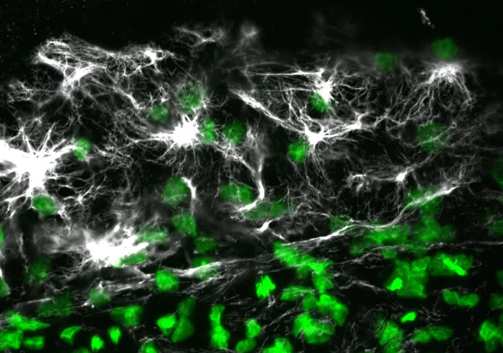
Avian embryos could join the list of model organisms used to study a specific type of cell migration called epiboly, thanks to the results of a study published this month in the journal Developmental Dynamics. The new study provides insights into the mechanisms of epiboly, a developmental process involving mass movement of cells as a sheet, which is linked with medical conditions that include wound healing and cancer.
The study, published online on March 15, explains how epithelial cells expand as a sheet and migrate to engulf the entire avian egg yolk as it grows. It also reveals the presence of certain molecules during this process that have not been previously reported in other major developmental models, including Xenopus frogs and zebrafish.
"These molecules and mechanisms of early development in the avian embryo may demonstrate evolutionary differences across species in the collective movement of epithelial cells and motivate additional studies of avian embryo development," said Evan Zamir, an assistant professor in the George W. Woodruff School of Mechanical Engineering at Georgia Tech.
Matt Futterman, who worked on the project as a graduate student at Georgia Tech, and mechanical engineering professor Andrés García also contributed to this study. The research was funded by Zamir's new faculty support from Georgia Tech and by a grant to García from the National Institutes of Health.
In the study, the researchers conducted immunofluorescence and high-resolution confocal microscopy experiments to examine the spatial distribution and expression of five proteins -- vimentin, cytokeratin, β-catenin, E-cadherin and laminin -- as cells moved to wrap the yolk sac of quail embryos during development.
The results showed that during this process, four of the proteins -- vimentin, cytokeratin, β-catenin and E-cadherin -- appeared in the cells located at the free edge of the migrating cell sheet. Finding dense interconnected networks of both vimentin and cytokeratin in the edge cells surprised the researchers.
"Since cytokeratin is generally associated with the epithelial phenotype and vimentin is generally associated with the mesenchymal phenotype, it's rare to see them expressed in the same cells, but this does occur in metastasizing tumor cells," said Zamir.
Cells expressing the mesenchymal phenotype are typically found in connective tissues -- such as bone, cartilage, and the lymphatic and circulatory systems -- whereas cells of the epithelial phenotype are found in cavities and glands and on surfaces throughout the body.
This finding provides evidence that epithelial cells normally attached to a membrane surface underwent biochemical changes that enabled them to assume a mesenchymal cell phenotype, which enhanced their migratory capacity. This process, called partial epithelial-to-mesenchymal transition, has many similarities to the initiation of tumor cell metastasis and wound healing.
Since this epithelial and mesenchymal expression pattern in the edge cells has not previously been reported in Xenopus or zebrafish, it may be unique to the avian embryo. This discovery would make the avian embryo a valuable model for studying tumor cell migration and wound healing.
In addition to detailing protein expression in the quail embryo during development, the researchers also determined the origin of the new cells required at the migrating edge to cover the growing yolk. During development, the radius of the quail yolk doubles every day for the first few days, representing a hundreds-fold increase in the egg yolk surface area.
"For each individual cell that has to cover the egg yolk as it grows, the migration around the yolk is extraordinary, because it's such a large territory -- it would be like an ant walking across the earth," explained Zamir.
Looking more closely at the edge cells, the researchers found strong evidence that expansion of the edge cell population was due exclusively to cells relocating from an interior region to the edge as the embryo expanded. The cells located at the free edge generated the bulk of the traction force necessary for expansion and towed the cells within the interior of the epithelium.
"These experiments confirm that edge cell proliferation is not the primary mechanism for expansion of the edge cell population," noted Zamir. "And our observation of epithelial-to-mesenchymal transition in the edge cells explains how these epithelial cells might be changing phenotype to become migratory in this rapidly expanding sheet."
To determine if this study's findings are indeed unique to the avian embryo, Zamir plans to conduct further studies to characterize protein expression and cell migration in Xenopus and zebrafish.
Research News & Publications Office
Georgia Institute of Technology
75 Fifth Street, N.W., Suite 314
Atlanta, Georgia 30308 USA
Media Relations Contacts: Abby Robinson (abby@innovate.gatech.edu; 404-385-3364) or John Toon (jtoon@gatech.edu; 404-894-6986)
Writer: Abby Robinson
News Contact
Abby Robinson
Research News and Publications
Contact Abby Robinson
404-385-3364
Mar. 09, 2011
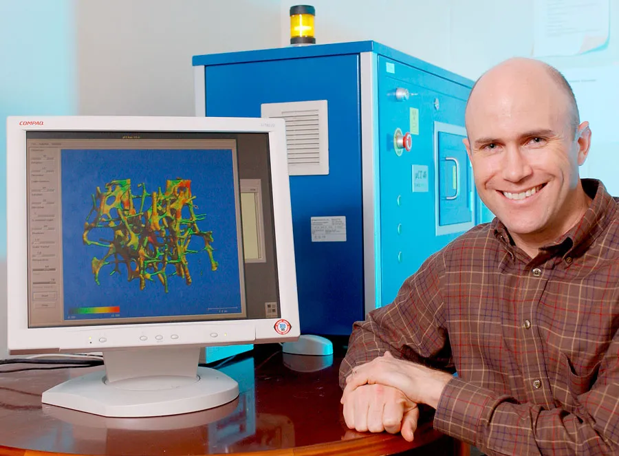
Robert E. Guldberg, director of Georgia Tech’sParker H. Petit Institute for Bioengineering and Bioscience (IBB), has beenappointed chairperson of the Musculoskeletal Tissue Engineering Study Sectionin the Center for Scientific Review – part of the National Institutes ofHealth.
Guldberg will serve as chairperson of the study section fromJuly 1, 2011, to June 30, 2013. The study section will contribute to thenational biomedical research effort and assure the quality of the NIH peerreview process.
Guldberg's research interests focus on musculoskeletalgrowth and development, functional regeneration following traumatic injury anddegenerative diseases, including skeletal fragility and arthritis
According to Dr. Toni Scarpa, director of the Center forScientific Review in NIH’s Department of Health and Human Services, Guldbergwas selected for the chair position because of his demonstrated achievement inhis scientific discipline, quality of research accomplishments, publications inscientific journals and overall judgment and objectivity.
At Georgia Tech, Guldberg studies cell-based therapies, bonebiomechanics, musculoskeletal injury, joint degeneration, biomaterials anddelivery, and micro-CT imaging. His laboratory creates strategies and enables technologiesfor the functional restoration of damaged or degenerated musculoskeletaltissues, with a focus on bone and cartilage.
In 1996, Guldberg joined Georgia Tech, serving both in IBBand the George W. Woodruff School of Mechanical Engineering. He was appointeddirector of IBB in November 2009.
Guldberg holds an undergraduate degree in mechanicalengineering, a master’s degree in bioengineering and mechanical engineering,and a Ph.D. in mechanical engineering from the University of Michigan.
News Contact
Georgia Tech Media Relations
Laura Diamond
laura.diamond@comm.gatech.edu
404-894-6016
Jason Maderer
maderer@gatech.edu
404-660-2926
Mar. 02, 2011
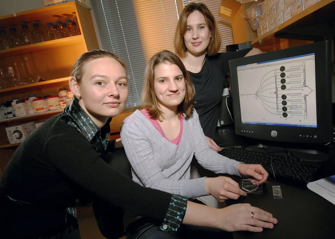
Manipulation of cells by a new microfluidic device may help clinicians improve a promising cancer therapy that harnesses the body's own immune cells to fight such diseases as metastatic melanoma, non-Hodgkin's lymphoma, chronic lymphocytic leukemia and neuroblastoma.
The therapy, known as adoptive T cell transfer, has shown encouraging results in clinical trials. This treatment involves removing disease-fighting immune cells called T cells from a cancer patient, multiplying them in the laboratory and then infusing them back into the patient's body to attack the cancer. The effectiveness of this therapy, however, is limited by the finite lifespan of T cells -- after many divisions, these cells become unresponsive and inactive.
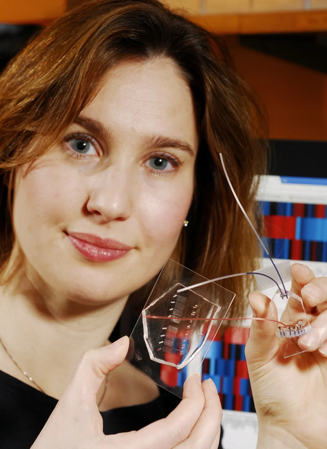
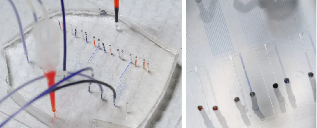
Researchers at Georgia Tech and Emory University have addressed this limitation by developing a microfluidic device for sample handling that allows a statistical model to be generated to evaluate cell responsiveness and accurately predict cell "age" and quality. Being able to assess the age and responsiveness of T cells -- and therefore transfer only young functional cells back into a cancer patient's body -- offers the potential to improve the therapeutic outcome of several cancers.
"The statistical model, enabled by the data generated with the microfluidic device, revealed an optimal combination of extracellular and intracellular proteins that accurately predict T cell age," said Melissa Kemp, an assistant professor in the Wallace H. Coulter Department of Biomedical Engineering at Georgia Tech and Emory University. "Knowing this information will help facilitate the clinical development of appropriate T cell expansion and selection protocols."
Details on the microfluidic device and statistical model were published in the March issue of the journal Molecular & Cellular Proteomics. This work was supported by the National Institutes of Health, Georgia Cancer Coalition, and Georgia Tech Integrative Biosystems Institute.
Currently, clinicians measure T cell age by using multiple assays that rely on measurements from large cell populations. The measurements determine if cells are exhibiting functions known to appear at different stages in the life cycle of a T cell.
"Since no one measurement is a perfect predictor, it is advantageous to concurrently sample multiple proteins at different time points, which we can do with our microfluidic device," explained Kemp, who is also a Georgia Cancer Coalition Distinguished Professor. "The wealth of information we get from our device for a small number of cells far exceeds a single measurement from a population the same size by another assay type."
For their study, Kemp, electrical engineering graduate student Catherine Rivet and biomedical engineering undergraduate student Abby Hill analyzed CD8+ T cells from healthy blood donors. They acquired information from 25 static biomarkers and 48 dynamic signaling measurements and found a combination of phenotypic markers and protein signaling dynamics -- including Lck, ERK, CD28 and CD27 -- to be the most useful in predicting cellular age.
To obtain biomarker and dynamic signaling event measurements, the researchers ran the donor T cells through a microfluidic device designed in collaboration with Hang Lu, an associate professor in the Georgia Tech School of Chemical & Biomolecular Engineering. After stimulating the cells, the device divided them into different channels corresponding to eight different time points, ranging from 30 seconds to seven minutes. Then they were divided again into populations that were chemically treated to halt the biochemical reactions at snapshots in time to build up a picture of the signaling events that occurred as the T cells responded to antigen.
"While donor-to-donor variability is a confounding factor in these types of experiments, the technological platform minimized the experimental data variance and allowed stimulation time to be precisely controlled," said Lu.
With the donor T cell data, the researchers developed a model to assess which biomarkers or dynamical signaling events best predicted the quality of T cell function. The model found the most informative data in predicting cellular age to be the initial changes in signaling dynamics.
"Although a combination of biomarker and dynamic signaling data provided the optimal model, our results suggest that signaling information alone can predict cellular age almost as well as the entire dataset," noted Kemp.
In the future, Kemp plans to use this approach of combining multiple cell-based experiments on a microfluidic chip to integrate single-cell information with population-averaged techniques, such as multiplexed immunoassays or mass spectrometry.
This project is supported in part by the National Institutes of Health (NIH)(Grant No. R21CA134299). The content is solely the responsibility of the principal investigator and does not necessarily represent the official views of the NIH.
Research News & Publications Office
Georgia Institute of Technology
75 Fifth Street, N.W., Suite 314
Atlanta, Georgia 30308 USA
Media Relations Contacts: Abby Robinson (abby@innovate.gatech.edu; 404-385-3364) or John Toon (jtoon@gatech.edu; 404-894-6986)
Writer: Abby Robinson
News Contact
Abby Robinson
Research News and Publications
Contact Abby Robinson
404-385-3364